

|Articles|November 1, 2006
Beauty of RCM more than skin deep
Author(s)John Jesitus
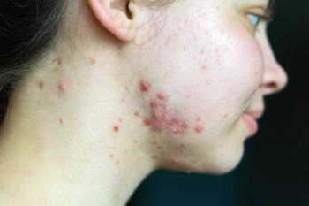
RCM has shown promise in clinical studies evaluating its potential in melanoma diagnosis.
Advertisement
RCM stands at the forefront of a handful of new technologies capable of offering in vivo, high-resolution methods for analyzing skin lesions, says Salvador Gonzalez, M.D., Ph.D., faculty/staff member of the dermatology service at Memorial Sloan-Kettering Cancer Center (MSKCC), New York.
Such technologies include optical coherence tomography, high-frequency ultrasound and magnetic resonance imaging (MRI).
More specifically, he says that in contrast to the minimum resolution of 10 μ to 15 μ that these technologies offer, "RCM provides the highest lateral resolution (less than 1 μ) and sectioning (3 μ to 5 μ) among all optical and nonoptical imaging modalities."
As such, he says RCM's resolution and sectioning capabilities are comparable to those provided by histology to a depth of 200 μm to 250 μm, which is sufficient for imaging the epidermis, papillary dermis and upper reticular dermis.
Furthermore, Dr. Gonzalez says that while routine pathology generally uses vertically oriented physical sections of skin, real-time RCM provides horizontal optical sections of the skin.
Promise for detection
Because skin cancers often spread laterally, Dr. Gonzalez tells Dermatology Times, "One may think that RCM offers a significant advantage compared to the standard vertical sections of histology."
Because RCM relies on naturally occurring optical stains such as melanin, which provides the strongest contrast in confocal reflectance images, Dr. Gonzalez says, "RCM is best suited for the in vivo examination of melanocytic lesions - benign and dysplastic nevi and melanomas - which it accomplishes with very high resolution and superb contrast."
Already, he reports that RCM has shown promise in clinical studies evaluating its potential in melanoma diagnosis.
"Based in part on the success of these European studies," he adds, "the National Cancer Institute recently has funded a large, multicenter clinical study to evaluate RCM for the screening and diagnosis of pigmented lesions."
Moreover, Dr. Gonzalez says, "We have shown - when I was at Harvard Medical School and now at MSKCC - that RCM may represent a promising adjunct tool to guide biopsies and avoid sampling errors resulting from clinical evaluation alone."
In this regard, Dr. Gonzalez says he and his colleagues have published a preliminary study showing RCM's effectiveness in presurgical margin mapping and margin demarcation of poorly defined malignant melanoma, lentigo maligna melanoma and also amelanotic melanoma (Arch Dermatol. 2004 Sep;140(9):1127-1132). More recently, he says, his team at MSKCC has undertaken a large-scale ongoing project to confirm this capability.
Additionally, he says researchers have systematically analyzed RCM's promise for screening and diagnosing other skin cancers - such as basal cell carcinoma (BCC), actinic keratoses, Bowen's disease and cutaneous T-cell lymphoma - relative to routine histology.
For example, Dr. Gonzalez says that regarding BCC, "In a retrospective multicenter clinical trial, we showed that the sensitivity and specificity of RCM in reference to routine histology was higher than clinical evaluation alone (J Am Acad Dermatol. 2004 Dec;51(6):923-930)."
Likewise, he says RCM's potential - which his group and others have proven feasible - to allow repeated evaluations of the same skin site over time holds promise for monitoring treatment response noninvasively and in vivo.
Advertisement
Related Content
Advertisement


Latest CME


Advertisement
Advertisement
Trending on Dermatology Times
1
FDA Accepts Addition of Bemotrizinol as First New Sunscreen Ingredient in 20 Years
2
AI Analysis Reveals Menopausal Hair Loss Linked to Systemic Health and Inflammation
3
Sun, Science, and Skin: Dermatology's Biggest Spotlights This June
4
Novel Botanical Moisturizer Demonstrates Superior Efficacy Over Metronidazole in Phase 2 Rosacea Trial
5