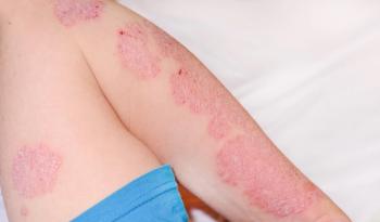
Atopic Dermatitis in Skin of Color
Key Takeaways
- AD presents differently in SOC, leading to potential misdiagnosis and delayed treatment, increasing morbidity.
- Clinical trials for AD lack diversity, with a majority of participants identifying as White.
A breakfast product theater at the 3rd Annual Society of Dermatology Nurse Practitioners Symposium, sponsored by Sanofi and Regeneron Pharmaceuticals, dove into the different manifestation of atopic dermatitis in skin of color and how to better recognize the disease in this patient population.
Atopic Dermatitis (AD) presents differently in patients with skin of color (SOC) vs lighter skin tones. This difference in presentation can be a cause of genetic factors, phenotypes, and histologic variations and can cause misdiagnosis in SOC patients. The way to combat this is to recognize clinical variation of AD in SOC and then develop strategies for this group of patients, according to a breakfast product theater at the 3rd Annual Society of Dermatology Nurse Practitioners Symposium, sponsored by Sanofi and Regeneron Pharmaceuticals.1
There are few studies that have investigated the epidemiological differences between SOC and lighter skin. In phase 2 and phase 3 clinical trials for AD, between the years of 2009 and July 2019, 54% of patients enrolled identified as White.2 Additionally, 8.9% identified as Black, 16% Asian, and 20.5% were other/unspecified. This demonstrates the lack of diversity in AD clinical trials, according to the session.
Even in medical school and residency, depending on where health care professionals are practicing, there can be a lack of knowledge on SOC. This can lead to delayed treatment for patients of color (POC) and increased morbidity or mortality from this delay. When compared to White patients, Black patients were 3 times more likely to be diagnosed with AD and Asian or Pacific Islanders were 7 times more likely.3
AD may also have a greater negative impact on quality of life (QOL) in SOC patients than their lighter skinned counterparts and Black patients had more physician visits for AD than White patients.4-6 Moreover, pediatric patients with SOC are more likely to have persistent AD and White patients are more likely to have controlled disease vs Black patients.7,8
Why could this be? It could be genetic variations like the Filaggrin (FLG) null mutations that could contribute to skin barrier dysfunction. This mutation can be different depending on the race of the patient.9 Patients would need to be tested to determine if a mutation is present and that does not always happen, according to the SDNP session. Additionally, Black patients are 6 times less likely to be tested for mutations compared to White patients.10
There could also be immunophenotypic variations between patients with AD of different races.
The way that AD presents itself is different in various skin tones and can be confusing for those who have not had much experience in darker skin types. Black patient may be at a higher risk of transepidermal water loss (TEWL) causing dry skin which has been studied in multiple small trials. This greater risk of dry skin would be consistent with a higher prevalence of AD.11
Beyond examining and making the right diagnosis of disease, many times there is an underestimation of AD severity in patients of SOC.11 This difficulty could be due to how erythema appears as dark purple in darker skin types. Missing this key symptom can lead to more severe disease.11-13
A common way to treat AD is topical cortisone steroids (TCS), but this treatment may lead to other skin issues such as hypopigmentation in SOC, especially in the Black population. This can dissipate with discontinuation of treatment, but does reveal a need for different treatment methods for SOC.12,14,15
When treating AD in patients with darker skin tones, key pearls to keep in mind include8,11,12:
- Long-term use of TCS may worsen hypopigmentation in darker skin types
- Higher doses of narrowband-UVB are needed to treat skin with darker pigmentation
- Delayed diagnosis and treatment can lead to greater severity in AD in SOC patients
- Self-identified Black patients may have worse disease control than White patients
- More data on the efficacy of treatments for AD in SOC population are needed
References:
- The shades of atopic dermatitis in skin of color. Sponsored by Sanofi and Regeneron Therapeutics. Presented at: The Society of Dermatology Nurse Practitioners Annual Symposium; April 22-23, 2022; Nashville, TN.
- Price KN, Krase JM, Loh TY, Hsiao JL, Shi VY. Racial and ethnic disparities in global atopic dermatitis clinical trials. Br J Dermatol. 2020;183(2):378-380. doi:10.1111/bjd.18938
- Shaw TE, Currie GP, Koudelka CW, Simpson EL. Eczema prevalence in the United States: data from the 2003 national survey of children’s health. J Invest Dermatol. 2011;131(1):67-73. doi:10.1038/jid.2010.251
- Wan J, Margolis DJ, Mitra N, Hoffstad OJ, Takeshita J. Racial and ethnic differences in atopic dermatitis–related school absences among us children. JAMA Dermatol. 2019;155(8):973. doi:10.1001/jamadermatol.2019.0597
- Poladian K, De Souza B, McMichael AJ. Atopic dermatitis in adolescents with skin of color. Cutis. 2019; 104(03):164-168.
- McGregor SP, Farhangian ME, Huang KE, Feldman SR. Treatment of atopic dermatitis in the United States: analysis of data from the national ambulatory medical care survey. J Drugs Dermatol. 2017;16(3):250-255.
- Kim Y, Blomberg M, Rifas-Shiman SL, et al. Racial/ethnic differences in incidence and persistence of childhood atopic dermatitis. J Invest Dermatol. 2019;139(4):827-834. doi:10.1016/j.jid.2018.10.029
- Abuabara K, You Y, Margolis DJ, Hoffmann TJ, Risch N, Jorgenson E. Genetic ancestry does not explain increased atopic dermatitis susceptibility or worse disease control among African American subjects in 2 large US cohorts. J Allergy Clin Immunol. 2020;145(1):192-198.e11. doi:10.1016/j.jaci.2019.06.044
- Park J, Jekarl DW, Kim Y, Kim J, Kim M, Park YM. Novel FLG null mutations in Korean patients with atopic dermatitis and comparison of the mutational spectra in Asian populations. J Dermatol. 2015;42(9):867-873. doi:10.1111/1346-8138.12935
- Margolis DJ, Apter AJ, Gupta J, et al. The persistence of atopic dermatitis and filaggrin (Flg) mutations in a US longitudinal cohort. J Allergy Clin Immunol. 2012;130(4):912-917. doi:10.1016/j.jaci.2012.07.008
- Vachiramon V, Tey HL, Thompson AE, Yosipovitch G. Atopic dermatitis in African American children: addressing unmet needs of a common disease. Pediatr Dermatol. 2012;29(4):395-402. doi:10.1111/j.1525-1470.2012.01740.x
- Kaufman BP, Guttman-Yassky E, Alexis AF. Atopic dermatitis in diverse racial and ethnic groups-Variations in epidemiology, genetics, clinical presentation and treatment. Exp Dermatol. 2018;27(4):340-357. doi:10.1111/exd.13514
- Ben-Gashir MA, Seed PT, Hay RJ. Quality of life and disease severity are correlated in children with atopic dermatitis. Br J Dermatol. 2004;150(2):284-290. doi:10.1111/j.1365-2133.2004.05776.x
- Eichenfield LF, Stein Gold LF. Addressing the immunopathogenesis of atopic dermatitis: advances in topical and systemic treatment. Semin Cutan Med Surg. 2017;36(2 Suppl 2):S45-S48. doi:10.12788/j.sder.2017.012
- Hengge UR, Ruzicka T, Schwartz RA, Cork MJ. Adverse effects of topical glucocorticosteroids. J Am Acad Dermatol. 2006;54(1):1-15; quiz 16-18. doi:10.1016/j.jaad.2005.01.010
Newsletter
Like what you’re reading? Subscribe to Dermatology Times for weekly updates on therapies, innovations, and real-world practice tips.


