
Publication|Articles|February 1, 2023
- Dermatology Times, May 2023 (Vol. 44. No. 05)
- Volume 44
- Issue 05
An Overview of Granulomatous Dermatitis
Granulomatous dermatitis includes a group of reactive dermatologic disorders characterized by distinct histopathological patterns, clinical manifestations, and associated diseases.
Advertisement
Granulomatous dermatitis (GD) includes a group of reactive dermatologic disorders characterized by distinct histopathological patterns, clinical manifestations, and associated diseases. The clinical and histologic subtypes encompassed by GD include palisaded neutrophilic and granulomatous dermatitis (PNGD) and interstitial granulomatous dermatitis (IGD).1 The etiology of these subtypes is not well understood. Because both disorders often occur in conjunction with systemic inflammation, it is thought that they are the result of immune dysregulation or dysfunction. Deposition of immune complexes in small dermal vessels in the context of a systemic disorder or due to an external exposure such as medication may lead to inflammation, collagen damage, and granulomatous infiltrates.2 This review will compare and contrast the features of each cutaneous eruption.
Palisaded neutrophilic granulomatous dermatitis (PNGD)
Clinical context:
The first of the granulomatous dermatitides includes PNGD, which may present as symmetric nodules and indurated plaques on the face and trunk, and/or umbilicated papules on the extensor surfaces of the extremities.2 Lesions may also occur as linear cords on the flank, often described as a “burning rope sign.” PNGD is thought to represent a cutaneous reaction pattern occurring in association with numerous systemic diseases, most commonly rheumatologic disorders such as connective tissue disease, inflammatory arthritides, and lymphoproliferative disorders.2 Others include inflammatory bowel disease, myelomas, systemic lupus erythematosus, malignancies, infection, and certain medications.3-5
Epidemiology:
Due to its rarity, little is known about the epidemiology of PNGD. While PNGD can occur in all ages, the disease is more rare in children. It affects females more than males in a ratio of approximately 3:1, which may reflect the distribution of underlying, associated systemic diseases. With regards to malignancy-associated PNGD, cutaneous manifestations may precede a malignancy diagnosis by up to five years.2
Diagnosis:
The diagnosis of PNGD is confirmed by skin biopsy. Histologic examination may vary based on lesion age, underlying etiologies, and associated diseases. While early lesions may reveal small-vessel leukocytoclastic vasculitis with a neutrophilic infiltrate, older lesions may show degenerated collagen and fibrosis. Palisading granulomas characteristic of PNGD may also be termed “Churg Strauss granulomas” or “flame figures.” Notably, a lack of mucin can help to distinguish PNGD from granuloma annulare.1-3,6 More recently, a unique histologic variant of PGND has been identified in patients with chronic myelomonocytic leukemia. In this variant, one may observe a wedge-shaped, dermal infiltrate with foamy multinucleated histiocytes and foci of necrobiotic collagen.7
Although the presentation is benign, the diagnosis of PNGD is important because the disease is often a cutaneous manifestation of a more serious systemic condition or underlying infection.
Management:
Patients presenting with any subtype of GD, including PGND, require an evaluation for potential triggers and associated systemic disorders. Medications that most commonly trigger PGND include calcium channel blockers, beta blockers, angiotensin converting enzyme inhibitors, and statins.6 A recent case report also highlights PGND with tocilizumab as the suspected trigger.8 Laboratory evaluation for potential autoimmune etiologies includes measurements for antinuclear antibody, antinuclear cytoplasmic antigen, rheumatoid factor, and citrullinated protein. Patients should also undergo age-appropriate cancer screening for malignancy-associated PGND.6
About 20% of patients show spontaneous resolution of PGND-associated skin lesions usually within a few weeks to a month.3
For the remaining 80%, treatment of PGND is primarily directed towards addressing the underlying disease or removing the inciting factor (for example, a medication). Treatments include topical corticosteroids, non-steroidal anti-inflammatory drugs, dapsone, colchicine, prednisone, oral tacrolimus, tumor necrosis factor (TNF) inhibitors, and baricitinib.8
Interstitial Granulomatous Dermatitis (IGD) and Interstitial Granulomatous Drug Reaction (IGDR)
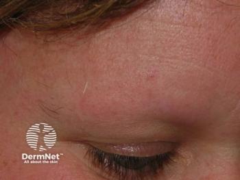
Interstitial granulomatous dermatitis (IGD) is another granulomatous dermatitis characterized by erythematous plaques and patches of various morphologies on the inner arms, thighs, trunk, and intertriginous areas. Other manifestations include macular erythema, subcutaneous nodules, and papules on the elbows. Although found in only 10% of patients, the presence of indurated, “rope-like” lesions that favor the lateral trunk and skin folds is pathognomonic for IGD.1
Similar to PGND, IGD often occurs in the setting of numerous systemic diseases, including rheumatological disorders, hematologic disorders, malignancies, and infections. Case reports of IGD induced by myelofibrosis and lymphomas illustrate the wide variety of associations.9 IGD can also be induced by medications; this condition is termed interstitial granulomatous drug reaction (IGDR) and is thought to be a distinct histopathologic entity.10 Clinical appearance of IGDR is similar to IGD with annular erythematous plaques on the proximal inner thighs, trunk, and intertriginous areas.3
Epidemiology:
Similar to PGND, the rarity of IGD/IGDR makes it difficult to study the epidemiology. As with PGND, IGD occurs in all ages but mostly in adults ages 40-50 and is also less common in children. IGD also tends to favor females over males in a 3:1 ratio. In the case of IGDR, onset of cutaneous lesions typically occurs months to years after starting the offending medication.10
Diagnosis:
IGD and IGDR are diagnosed by histology on a skin biopsy. Histopathological findings of IGD include palisading granulomas composed of histiocytes around foci of degenerated collagen and a dermal interstitial lymphocytic infiltrate composed of neutrophils. Other distinct features include a lack of mucin and unlike PNGD, a lack of vasculitis.6 Some histopathological findings of IGDR overlap with IGD such as interstitial histiocytes surrounding degenerating collagen. Unique features of IGDR include lymphoid atypia, vacuolar interface dermatitis with dyskeratosis, and prominent eosinophilia.2,6,10
Management:
Like PGND, patients with IGD require an evaluation for potential triggers and associated systemic disorders. Medications associated with IGDR include beta blockers, calcium channel blockers, angiotensin-converting enzyme inhibitors, statins, furosemide, antidepressants, and anticonvulsants. Case reports have also implicated hydrochlorothiazide, ustekinumab, and tocilizumab.4 Interestingly, TNF-alpha inhibitors and ustekinumab, despite being used to treat IGD, have paradoxically also caused IGDR.12,13
Treatments for IGD include cessation of the inciting agent and/or addressing the underlying associated systemic condition. Topical corticosteroids, intralesional corticosteroids, dapsone, hydroxychloroquine, and ustekinumab have been shown to improve cutaneous symptoms.13
What’s New in Granulomatous Dermatitis:
As described above, there is significant overlap in clinical presentation and context of PNGD and IGDR. Although there are various distinct histological and clinical patterns that appear to differentiate the two, this distinction may be insignificant. Numerous recent reports of granulomatous reactions to anti-cancer therapies that do not clearly fall into either disease category have further blurred the distinction between PNGD and IGDR. Due to similarities in clinical manifestations, treatment, and overall clinical course, recent literature has advocated for the grouping of PNGD and IGDR under the umbrella term “reactive granulomatous dermatitis”.6
References
1. Rodríguez-Garijo N, Bielsa I, Mascaró Jr J m., et al. Reactive granulomatous dermatitis as a histological pattern including manifestations of interstitial granulomatous dermatitis and palisaded neutrophilic and granulomatous dermatitis: a study of 52 patients. Journal of the European Academy of Dermatology and Venereology. 2021;35(4):988-994. doi:10.1111/jdv.17010
2. Imadojemu S, Rosenbach M. Advances in Inflammatory Granulomatous Skin Diseases. Dermatologic Clinics. 2019;37(1):49-64. doi:10.1016/j.det.2018.08.001
3. Stiff KM, Cohen PR. Palisaded Granulomatous Dermatitis Associated with Ulcerative Colitis: A Comprehensive Literature Review. Cureus. 9(1):e958. doi:10.7759/cureus.958
4. Zabihi-pour D, Bahrani B, Assaad D, Yeung J. Palisaded neutrophilic and granulomatous dermatitis following a long-standing monoclonal gammopathy: A case report. SAGE Open Med Case Rep. 2021;9:2050313X20979560. doi:10.1177/2050313X20979560
5. Akagawa M, Hattori Y, Mizutani Y, Shu E, Miyazaki T, Seishima M. Palisaded Neutrophilic and Granulomatous Dermatitis in a Patient with Granulomatosis with Polyangiitis. CDE. 2020;12(1):52-56. doi:10.1159/000506670
6. Wanat KA, Caplan A, Messenger E, English JC, Rosenbach M. Reactive granulomatous dermatitis: A useful and encompassing term. JAAD Int. 2022;7:126-128. doi:10.1016/j.jdin.2022.03.004
7. Enescu CD, Patel A, Friedman BJ. Unique Recognizable Histopathologic Variant of Palisaded Neutrophilic and Granulomatous Dermatitis that Is Associated With SRSF2-Mutated Chronic Myelomonocytic Leukemia: Case Report and Review of the Literature. Am J Dermatopathol. 2022;44(3):e33-e36. doi:10.1097/DAD.0000000000002085
8. Hung YT, Chung WH, Chen CB, Chan TM. Baricitinib Treatment for Palisaded Neutrophilic Granulomatous Dermatitis: A New Paradoxical Reaction to Tocilizumab [published online ahead of print, 2022 May 12]. Dermatitis. 2022;10.1097/DER.0000000000000879. doi:10.1097/DER.0000000000000879
9. Rose C, Holl-Ulrich K. [Granulomatous reaction pattern of the skin : Interstitial granulomatous dermatitis - lymphoma - vasculitis]. Hautarzt. 2017;68(7):553-559. doi:10.1007/s00105-017-4004-6
10. Aldibane RT, Hawsawi KA. Interstitial Granulomatous Drug Reaction: A Case Report. Cureus. 2022;14(2). doi:10.7759/cureus.21893
11. Altemir A, Iglesias-Sancho M, Sola-Casas M de los Á, Novoa-Lamazares L, Fernández-Figueras M, Salleras-Redonnet M. Interstitial granulomatous dermatitis following tocilizumab, a paradoxical reaction? Dermatologic Therapy. 2020;33(6):e14207. doi:10.1111/dth.14207
12. Altemir A, Iglesias-Sancho M, Sola-Casas M de los Á, Novoa-Lamazares L, Fernández-Figueras M, Salleras-Redonnet M. Interstitial granulomatous dermatitis following tocilizumab, a paradoxical reaction? Dermatologic Therapy. 2020;33(6):e14207. doi:10.1111/dth.14207
13. Walker A, Westerdahl JS, Zussman J, Mathis J. Interstitial Granulomatous Drug Reaction to Ustekinumab. Case Rep Dermatol Med. 2022;2022:1461145. doi:10.1155/2022/1461145
14. Cover image: Ahmed Z, Joad S, Singh, et al. Interstitial granulomatous dermatitis successfully treated with etanercept. Am J Case Rep, 2014; 15: 94-96 DOI: 10.12659/AJCR.890074
Articles in this issue
about 3 years ago
Discovering Dermatology Times: May 2023about 3 years ago
Consent and Experienceabout 3 years ago
Best Practices for Identifying and Managing Granulomatous Dermatitisabout 3 years ago
An Overview of Acral Peeling Skin Syndromeabout 3 years ago
Managing Keloids and Hypertrophic Scarsabout 3 years ago
Expanding Ethnic Hair Careabout 3 years ago
Alopecia’s Impact on Mental Healthabout 3 years ago
Exposure to Heavy Traffic Affects Risk of Atopic Dermatitisabout 3 years ago
Preparing Patients for the Summer Sun: Location, Location, LocationAdvertisement
Related Content
Advertisement


Latest CME


Advertisement
Advertisement
Trending on Dermatology Times
1
FDA Approves Roflumilast Cream 0.3% for Plaque Psoriasis Down to Age 2
2
Evommune's EVO756 Misses Primary End Point in Phase 2b CSU Trial, Development Continues in AD and Migraine
3
Dermatology Times June 2026 Recap
4
Why Joint Damage Inhibition Matters in Psoriatic Arthritis Treatment
5