

|Articles|December 18, 2020
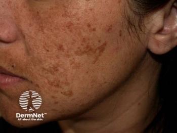
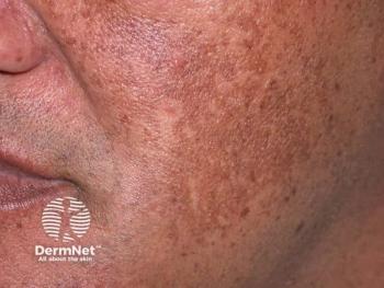
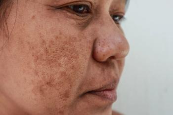
Targeting melasma’s vascular component improves treatment outcomes, prevent relapse
Author(s)Lisette Hilton
Dermatologists should consider assessing melasma patients for increased vascularity and include antivascular treatments to improve outcomes among patients whose lesions have a vascular component.
Advertisement
Dermatologists should consider assessing melasma patients for increased vascularity and include antivascular treatments, including tranexamic acid and laser and light therapies, to improve outcomes among melasma patients whose lesions have a vascular component.
“Methods of evaluation of the vascular component of melasma are relatively accessible and may positively impact the patient’s clinical course, particularly for recalcitrant melasma cases,” researchers from the Department of Dermatology at State University of New York reported in their study “
In a systematic review of 34 published research articles, the authors found strong evidence for what they called “the important and underreported vascular component of melasma.”
Melasma treatment remains challenging despite conventional melasma treatment regimens focused on sun protection and lightening agents. Genomics, hormonal imbalance and UV exposure are widely accepted contributors to melasma pathogenesis. But evidence is building that suggests there may be an important vascular component to melasma pathogenesis, according to the paper.
“Emerging evidence indicates that melasma pathogenesis may involve aberrant angiogenesis and vasodilation of dermal blood vessels, rather than solely increased melanogenesis resulting in hyperpigmentation,” the authors wrote.
The authors hypothesize that increased vasculature, vasodilation and inflammation result dermal capillaries in melasma lesions becoming increasingly permeable. The capillaries leak red blood cells, which contributes to hyperpigmentation that might come from solar light or light from electronic devices.
The evidence
Histological evaluations and studies looking at proangiogenic and vascular marker expressions seem to support that melasma has a vascular component. Included among those are several studies looking at melasma biopsy samples, which show melasma lesions have increased vascularity compared to normal skin.
Authors of a diagnostic study found vascular endothelial growth factor (VEGF) is upregulated in melasma but did not find that VEGF was related to melanin synthesis, melanocyte proliferation or melanocyte migration.
Researchers have also reported an overexpression of inducible nitric oxide synthase (iNOS), a vasodilation mediator, is featured in melasma lesions. Adding to the evidence, researchers have found endothelial cells play an important role in physiologic pigmentation, but more research is needed to characterize that role in melasma, according to the paper.
Studies looking at clinical evidence of vascular characteristics in melasma have found visibly increased vascularity in melasma lesions, including distinct telangiectatic erythema.
Researchers have used dermoscopy in diagnostic studies to reveal melasma hypervascularity. Those vascular features improved with treatment, according to the paper.
“Patients with visibly widened capillaries on dermoscopy showed greater improvements in the Melasma Area and Severity Index (MASI) score after several antivascular treatments, including tranexamic acid (TXA) and combination Q-switched Nd:YAG (QS Nd:YAG) laser and pulsed dye laser (PDL), compared with patients without these dermoscopic features,” they wrote.
Which antivascular treatments appear to work?
The authors reviewed five studies, including case reports, on outcomes using the 595nm pulsed dye laser to treat melasma. Researchers have found that pulsed dye laser treatment, which destroys blood vessels by targeting the chromophore oxyhemoglobin, improves melasma’s vascularity.
Combinations seem to work well. In a split-face study, daily treatment with the pulsed dye laser along with triple combination therapy with hydroquinone, tretinoin and fluocinolone led to a significant and sustained decrease in the MASI score. In one patient who presented three years later with a melasma relapse, the lesions were not in the previously treated areas.
Authors of a retrospective review studied the efficacy of melasma treatment combining pulsed dye laser with a 1927 nm low-power fractional diode laser and found 54% of patients had great than 50% improvement in the presentation of their melasma compared to baseline.
Even the 1064 nm Q-switched Nd:YAG laser was clinically effective as a melasma treatment in three studies. But researchers looking at melasma treatment with the copper bromide laser have reported conflicting results, according to the paper.
The authors pointed out that pulsed dye laser, Q-switched Nd:YAG and other light therapies can result in post-inflammatory hyperpigmentation and other adverse events, particularly in patients with darker skin types.
Researchers have reported improvements in melasma after treatment with the pharmacologic therapy tranexamic acid, whether given orally or topically. In one study, patients with vascular dermoscopic features had greater improvement after applying a tranexamic acid topical for 8 weeks than those who applied a cream without tranexamic acid. In vitro research suggests tranexamic acid decreases expression of several VEGF receptors in endothelial cells and melanocytes, as well as decreases melanocyte proliferation, melanin content, and melanogenic protein expression, according to the authors. Other research suggests tranexamic acid might decrease melasma vascularization by vasoconstriction.
What should dermatologist to do?
Melasma remains a therapeutic challenge, and dermatologists have easy access to ways in which they can assess and treat a possible vascular component to improve outcomes.
In addition to physical examination for evaluating melasma lesion vascularity, dermatologists might use dermoscopy, colorimetry and reflectance confocal microscopy.
“After evaluation of vascularity, dermatologists may then consider adding targeted antivascular therapy to the patient’s treatment regimen,” they wrote.
While the therapeutic options include laser and light therapies and tranexamic acid, the authors recommended that dermatologists use an antivascular therapeutic based on patient preference, treatment goals and therapeutic availability. Dermatologists should weigh treatment benefits with risks, they wrote.
Future research on the topic should include randomized controlled trials, larger sample sizes, longer follow up and standardized evaluation methods, according to the paper.
Reference:
1 Masub N, Nguyen JK, Austin E, Jagdeo J. The Vascular Component of Melasma: A Systematic Review of Laboratory, Diagnostic, and Therapeutic Evidence. Dermatol Surg. 2020 Dec;46(12):1642-1650. doi: 10.1097/DSS.0000000000002770. PMID: 33252894.
Disclosures: None reported.
Advertisement
Related Content
Advertisement

Latest CME


Advertisement
Advertisement
Trending on Dermatology Times
1
Social Media Mythbusters: The Summer Skin Care "Shutdown"
2
Zasocitinib Shows High Skin Clearance at Hard-to-Treat Psoriasis Sites
3
New Review Confirms How Nonmedicated Skin Care Vehicles Drive Clinical Improvement
4
Breakout Bulletin: July 13-July 17
5