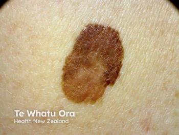
Suspected pediatric melanomas should be biopsied as they would be in adults
Pediatric melanomas are uncommon, but because melanoma is not always as easily diagnosed in children as in adults, it is critical to perform biopsies for pediatric patients just as one would for adults, an expert says.
Key Points
"Anything you would biopsy on an adult, should be biopsied on a child," says Ronald Hansen, M.D., professor of medicine, pediatrics and dermatology, University of Arizona, and medical staff section chief, Phoenix Children's Hospital.
While it may seem that the incidence of childhood melanoma is on the upswing, Dr. Hansen says it's not definitive whether it actually occurs more often, or if it's simply being reported more frequently.
Unique presentation
Pediatric melanoma presents very differently than melanomas generally seen in adults, where the vast majority are sun-induced, Dr. Hansen says.
"Those start with a broad, multicolored flat lesion we call superficial spreading melanomas, which aren't common in kids until they pass the age of about 15. That's when we start to see adult-type melanomas," he says.
Melanomas in children tend to be nodular lesions that grow thick, rather than broad, and represent nearly half of all pediatric melanomas.
"In that group, you have a number of nodular melanomas, but the Spitz tumors tend to be the dominant ones. Those are tricky to diagnose because they grow so rapidly."
Furthermore, nodular melanomas present a particular challenge in pediatric patients, Dr. Hansen says.
"They are harder to recognize because everything we learned about the ABCDs of diagnoses of melanomas - bigger than 6 mm, irregular borders, great color variability and funny shapes - don't necessarily apply in nodular melanoma," he says. "Nodular melanomas can be rapidly growing with black, red or brown lumps. Small ones are papules; big ones are nodular. Often, it's not until they break down and form a scab, a crust or an ulcer that someone recognizes that something's wrong. That's very late in the disease process.
"By the time that occurs, the melanomas are usually approaching 2 mm in depth or more, and are usually high-risk and are frequently fatal, metastasizing lesions in both children and adults."
Diagnostic options
Children appear to have a better survival rate with deep melanomas than do adults, Dr. Hansen says.
"That's part of the reason for controversy over whether some of the thick, atypical moles, such as the Spitz tumors, are melanomas at all, even though they look like melanomas under the microscope," he says. "That has led to speculation we may need something better than microscopic examination for diagnosing these lesions."
Genetic tests that can help to confirm a melanoma diagnosis are under investigation, according to Dr. Hansen.
"Gene tests, such as fish studies, are not readily available everywhere, and are not necessarily the perfect test," he says, "but they may add a dimension beyond just an expert pathologist determining that these are high-risk lesions."
The second-most common childhood melanoma occurs in congenital nevi. Dr. Hansen says they can be relatively common in giant nevi, which measure more than 20 cm across.
"Those have a 5 percent chance of developing melanoma in the child's lifetime, with the highest risk occurring in the first 10-12 years of life," he says. "That risk compares to perhaps a one in 1,000 or one in 10,000 risk of melanoma in a small congenital nevus, measuring 2-3 cm in size. The risk in nevi in between have an intermediate risk."
The problem, Dr. Hansen says, is that giant nevi are so large, they are often impossible to remove surgically. Smaller ones can be removed more easily, but carry little risk.
Lesion removal
Melanomas are easier to spot on these smaller lesions, he says, because they are a flat, single color, and parents are told to watch for focal area color change, such as a brown/black mole developing a focal area of gray, blue, red or white elevated papules.
The location of congenital nevi also plays a role in the risk of pediatric melanoma, Dr. Hansen says. If they occur over the extremities, the risk is lower. But nevi occurring on the back, especially over the spine, carry a higher risk.
"If I saw an intermediate-sized nevus over the spine, I might elect to have them removed, because there could be some prevention in doing that," he says.
The only treatment for childhood melanoma is early diagnosis and removal. Prophylactic removal of congenital nevi of all sizes is controversial, but the only curative treatment is surgical removal.
"Although promising new treatments for metastatic melanoma are being studied for adults, they haven't been tried in children yet," Dr. Hansen says. "The only time chemotherapy or radiation is used, is if cancer has spread to the lymph nodes. These treatments don't work very well, so if the melanoma is metastatic, chances of survival are very low."
Disclosures: Dr. Hansen reports no relevant financial interests.
Newsletter
Like what you’re reading? Subscribe to Dermatology Times for weekly updates on therapies, innovations, and real-world practice tips.


