

News|Articles|August 11, 2023
Study Shows Lupus Band Test Can Be Useful In Diagnosing Lupus Erythematosus
Author(s)Heather Raglin, Editor
Investigators wanted to find a testing option if other parameters are negative or conflicting and LE is suspected.
Advertisement
Researchers sought to assess the sensitivity and specificity of the lupus band test (LBT) in diagnosing lupus erythematosus (LE) following clinico-pathological correlation (CPC). Their findings showed that although the LBT is not a sensitive diagnostic tool for LE, it is highly specific and can be considered as a supportive tool in diagnosing LE.1
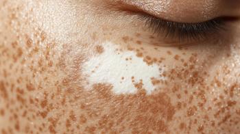
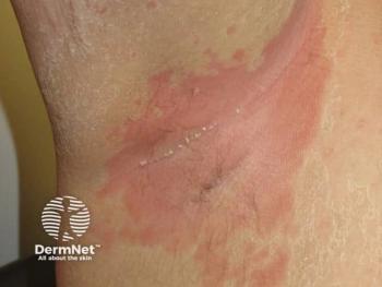
Of patients diagnosed with systemic lupus erythematosus (SLE), 85% will have a cutaneous manifestation of LE, most often photo-distributed erythema. CPC is necessary because the histological features of the subsets of cutaneous lupus erythematosus (CLE) overlap and vary with disease severity.
“The LBT is positive in lesional skin of 50%–94% of patients with SLE, 60% of patients with subacute CLE (SCLE) and 60–80% of patients with discoid LE (DLE).”1 Sun-exposed skin has a higher rate of positive LBTs (60%-70%) than skin not exposed to the sun (50%-60%). Biopsies of sun-exposed normal skin of young adults who do not have LE may have positive LBT 20% of the time.
To confirm a diagnosis of LE, direct immunofluorescence (DIF) may be used in addition to histology. Researchers evaluated biopsies sent to the pathology department for DIF between 2011 and 2018. A linear band deposition of immunoglobulins at the basal membrane (BM) was found in all biopsies that were positive for the LBT.
Clinical records of patients were reviewed for CPC and to determine if a definitive diagnosis of LE was made following CPC. Sensitivity and specificity for the LBT, histology, and serology were calculated using diagnosis following CPC as the gold standard.
DIF was requested on 947 biopsies, of which 27% (256/947) had a clinical query that included LE. Of these, 43.4% suspected LE as the main diagnosis, and 56.6% had a list of possible diagnoses, of which LE was 1. Mean patient age was 55 years and 75% were female. Of all the DIF biopsies received, sun-exposed sites, including the face, hands, and neck, accounted for 39.8%, and for 52.2% of biopsies with a positive LBT.
Of the 256 samples with a clinical query including LE, 9% had a positive LBT. LE was diagnosed in 39.8% of skin biopsies on histology, of which 18.6% had a positive LBT. “Only 37% (95/256) of patients had serology tested for autoantibodies. All 95 samples were tested for ANA [antinuclear antibodies], of which 60% (n = 57) were positive. LE-specific antibodies (dsDNA, anti-Sm, Ro/SS-A, La/SS-B) were tested in 48.4% (n = 46) of those who had ANA testing. Positive dsDNA was seen in 5.3% (5/95), positive anti-Sm in 3.2% (3/95), positive Ro/SS-A in 31.6% (30/95) and positive La/SS-B in 17.9% (17/95).”1
In 62.5% (160/256) of cases, a definitive diagnosis was made after CPC. Of those, LE was diagnosed in 46.3% (74/160).
In 102 of the 256 cases (39.8%), LE was diagnosed on histology, and of those, 18.6% (19/102) had a positive LBT. A positive LBT was found in 9% (23/256) of all biopsies for DIF. In 82.6% (19/23) of those, histological features were consistent with LE. “Autoantibody testing was performed in 12 of those with a positive LBT, of which 83.3% (10/12) were positive for ANA alone or in association with dsDNA, Ro/SS-A, and La/SS-B.”1 Of all cases with a positive LBT, 87% (20/23) had either clinical diagnosis of LE, histology consistent with LE, and/or serology consistent with LE.
All cases with a positive LBT had a homogenous thready or granular band of immunoglobulins and complement at the BM.
Of the 256 samples sent for DIF with suspicion of LE, 91% (233) had a negative LBT. Of those, 7% (17/233) had serological, histological, and clinical features consistent with LE. “Overall, 46.3% (109/233) with a negative LBT had either one, two or three of the following: clinical diagnosis of LE, histology consistent with LE, serology consistent with LE.”1
The sensitivity for diagnosing LE with the LBT was 17.6% and the specificity for diagnosing LE with the LBT was 98.8%. Diagnosing LE on histology alone had a sensitivity of 86.5% and a specificity of 90.7%.
The authors concluded that the LBT can be a useful tool for diagnosing LE when other parameters are negative or conflicting and LE is suspected.
Reference
- Ní Maolcatha S, Nic Dhonncha E, O’Connor C, Dinneen S, Heffron CCBB. The lupus band test: a review of the sensitivity and specificity in the diagnosis of lupus erythematosus. Skin Health Dis. 2023;3(4):e205.
https://doi.org/10.1002/ski2.205
Advertisement
Related Content
Advertisement


Latest CME


Advertisement
Advertisement
Trending on Dermatology Times
1
Bempikibart Shows Positive Phase 2a Results in Alopecia Areata
2
Study Suggests Non-Ablative Fractional Laser Reverses Epigenetic Signatures of Skin Aging
3
Roundtable Discussion Weighs Access and Adherence Challenges in Vitiligo Care
4
The Gut-Skin Axis: New Evidence Links Microbiota to Skin Disease via Inflammatory Cytokines
5