
|Articles|February 2, 2015
Study examines melanoma cells’ influence on keratinocytes
Results from a study on the effect of melanoma cells (MC) on human primary keratinocytes (HPK) has been released. Checkout the findings
Advertisement
A team of researchers from the Czech Republic has released the results of their study on the effect of
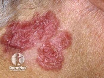
Noting that nodular melanoma is characterized by relatively poor therapeutic outcomes, the researchers started with the premise that, as with other tumors, “a permissive microenvironment is essential for melanoma progression.” They write that the presence of MC is as an important modulator of the tumor microenvironment, comparing it to the effect of nonmalignant cells also originating from neural crest (NCSC).
After morphometrical and immunohistochemical analyses of epidermis surrounding nodular melanoma were performed, the data were compared to results of transcriptome profiling of in vitro models, in which HPK were co-cultured with MC, normal human melanocytes and NCSC, respectively. Differentially expressed candidate genes were verified, and biological activity of candidate proteins was assessed on cultured HPK.
The researchers found that in 90 percent of the cases, the epidermis surrounding nodular melanoma exhibited hyperplastic features, which exhibited aberrant suprabasal expression of keratin 14 and loss of keratin 10. They found that MC and NCSC can increase expression of keratins 8, 14, 19 and vimentin in the co-cultured HPK, and note that this in vitro finding partially correlates with pseudoepitheliomatous hyperplasia observed in melanoma biopsies. The researchers also observed evidence of basic fibroblast growth factor (FGF-2), chemokine ligand (CXCL-1), interleukin-8 (IL-8) and vascular endothelial growth factor (VEGF-A) participation in the activity of melanoma cells on keratinocytes.
“Our data support the hypothesis of the importance of the microenvironment in melanoma biology, but our understanding of these mechanisms is still limited.” study author Ondrej Kodet, M.D., of Charles University in Prague, Czech Republic, tells Dermatology Times. “Mutual intercellular interactions in melanoma seem to participate in formation of this tumorigenic microenvironment. Our work is an experimental study; the results haven’t brought new data for clinical use yet, but these findings bring a new understanding of the complexity of tumor biology.”
Kodet O, Lacina L, KrejÄí E, et al.
More Melanoma articles
Advertisement
Related Content
Advertisement


Latest CME


Advertisement
Advertisement
Trending on Dermatology Times
1
What Dermatologists Need to Know About the First New Sunscreen Ingredient in Decades
2
The Expanding Role of Immunotherapies for Non-Melanoma Skin Cancers
3
Social Media Mythbusters: Skin Cycling
4
Filling the HS Treatment Gap: Ruxolitinib Targets Early-Stage Disease
5