Staging, developing parameters important before CTCL therapy
CTCL tumors can arise in patients with MF or in those with non-MF CTCL, who lack the typical MF signs of scaly patches or lesions. Almost all CTCL tumors present as bumps or ulcers.
"While most patients have the classic form of the disease - mycosis fungoides - there are also a significant number of variations," explains Peter W. Heald, M.D., professor of dermatology at the Yale University School of Medicine in New Haven, Conn. The variations of CTCL fall into different categories, including the clinical variants, biopsy variants and cellular or molecular variants.
Variants
A second categorization of CTCL variants consists of those defined by biopsy. These involve a distinctive finding on biopsy, such as pagetoid reticulosis, that leads to the classification. The purpura-type variant is associated with blood vessel leakage, which leads to bleeding in skin, while the granulomatous variant is associated with the development of granulomas.
As with clinical variants, clinical and pathologic features are incorporated in determining the exact biopsy variant involved. Fortunately, these findings are so distinct that classifying a CTCL is a reproducible task at the varied clinics dealing with the disease. Reproducibility is a key component to any classification scheme.
In addition to the clinical and biopsy-related variants, a third group of variants is identified and classified using special cellular and molecular studies performed on biopsy samples. Most of these are carried out using immunoperoxidase assays.
Dr. Heald explains, "We may find that the patient has a CD8+ suppressor cell lymphoma instead of the usual CD4+ helper cell lymphoma. Alternatively, patients may have involvement of gamma delta-T cells or, even more rarely, NK T cells." These less common CTCLs would be discovered in the process of using these special stains.
Erythrodermic variants of CTCL
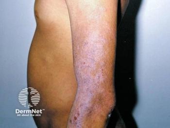
In the case of patients who present with generalized erythroderma and no discrete lesions, the bulk of the diagnosis relies upon blood tests, although some clinical features can suggest lymphoma. These include the "deck chair sign," in which skin is generally red with areas of sparing, particularly in skin folds. These patients also tend to develop a lower lid droop and thick, scaly skin on the palms and soles.
Dr. Heald notes that, "For these patients with erythroderma the diagnosis of CTCL is difficult to make on biopsy, but is readily made with blood work. The key is having someone look at the blood smear to examine the morphology of lymphocytes."
Flow cytometric analysis is also crucial, as the most reliable sign of CTCL in most patients with lymphoma-related erythroderma is an elevated (over six) CD4:CD8 ratio. This analysis will differentiate CTCL from psoriasis and eczema.
Several types of erythroderma-associated CTCL have been identified. Sezary syndrome refers to a spontaneous development of redness with no prior lesions or MF diagnosis. These patients have redness over 80 percent of their bodies, atypical blood cells on their smears and some other evidence of disease, such as flow cytometric or molecular findings. Sezary syndrome contrasts with erythrodermic MF, a disease progression from a patient's MF patches, plaques or tumors. The third type, erythrodermic CTCL, includes patients whose symptoms are not covered under the other two definitions - Either they do not have 80 percent body surface area coverage or they lack atypical cells on a blood smear.
CTCL-associated tumors
CTCL tumors can arise in patients with MF or in those with non-MF CTCL, who lack the typical MF signs of scaly patches or lesions. Almost all CTCL tumors present as bumps or ulcers.
Newsletter
Like what you’re reading? Subscribe to Dermatology Times for weekly updates on therapies, innovations, and real-world practice tips.


