

|Articles|April 1, 2013
Staged excision techniques for lentigo maligna address challenges
The many vagaries of lentigo maligna (LM) or melanoma in situ typically demand staged excision techniques for maximal margin control, according to an expert who spoke at the 71st annual American Academy of Dermatology conference.
Advertisement
Miami Beach, Fla. - The many vagaries of lentigo maligna (LM) or melanoma in situ typically demand staged excision techniques for maximal margin control, according to an expert who spoke at the 71st annual American Academy of Dermatology conference.
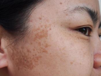
Because LMs occur in sun-damaged areas, says Kishwer S. Nehal, M.D., “They present in a very gradual, subtle way” that can be difficult for patients to notice, and for doctors to diagnose. She is director of Mohs surgery and dermatologic surgery, Memorial Sloan-Kettering Cancer Center, New York.
Additionally, “LMs can often be fairly large lesions.” This creates problems regarding biopsies and treatment. In the former area, she explains, it’s difficult to completely biopsy a facial lesion measuring one to two cm without leaving a large defect.
“Because they’re such large lesions, often only a portion of the melanoma has been biopsied before these patients are referred to surgery. When only a partial biopsy is feasible, the most suspicious area clinically or dermoscopically should be selected,” she says.
Treating such lesions creates challenges given location in functionally and cosmetically sensitive areas such as the nose, eyelids or ears, she adds.
More than meets the eye
“The other issue with the management of LM is that there can be much more underneath the skin than meets the eye. The standard melanoma treatment guidelines say to use 5 mm margins for an in situ lesion and one cm if it’s invasive (lentigo maligna melanoma/LMM). But up to half the time, those margins do not work in this subset of melanomas because LM has very unpredictable subclinical extension,” Dr. Nehal says. “You can’t just go by the pigment on the skin to determine how much margin to take. Woods light or mapping biopsies can be helpful in better delineating LM margins.”
Standard wide excision of LM results in recurrence rates between 8 and 20 percent, Dr. Nehal says. As an alternative, she says, surgeons frequently use Mohs surgery or staged excision techniques for LM and LMM.
“The obvious advantage of Mohs surgery is that it allows you to map the margins, and within one to two hours get a frozen section diagnosis at the margin. There is some difficulty, however, in interpreting atypical melanocytes relative to the surrounding sun-damaged skin.” Therefore, she says, some Mohs surgeons use immunostains to highlight the melanoma cells.
Staged excision
Conversely, “Our group at MSKCC uses staged excision, which is essentially a variation on Mohs surgery.” For LM, surgeons start by marking a standard 5 mm excision margin around the visibly pigmented lesion or biopsy site and maintaining orientation with marks at 12, 3, 6 and 9 o’clock, she says.
Next, using local anesthesia, the surgeons debulk the tumor by excising the residual clinical lesion as a full-thickness disk to the deep subcutis, given frequent follicular involvement. This disc goes into a formalin bottle for histologic analysis and melanoma staging.
Surgeons subsequently excise margins to the deep subcutis and send each tissue quadrant to pathology (in a separate bottle) with a suture marking to preserve orientation. To keep track of orientation on the patient, surgeons place small scores at the edge of the defect at the 12, 3, 6 and 9 o’clock sites.
Excised tissue is processed rush with paraffin-embedded vertical sections at close intervals. The next day, patients return for re-excision of any incompletely excised margins, and the above process is repeated until clear histologic margins are achieved (Hazan C, Dusza SW, Delgado R, et al. J Am Acad Dermatol. 2008;58(1):142-148). In this regard, “A 2 to 3 mm histologic margin of clearance is preferred to reduce risk of local recurrence.” A control biopsy from normal sun-damaged skin can help histologically differentiate residual melanoma in situ from atypical melanocytes at margins, she adds.
“The main reason we don’t close the wound until margins are clean is that many of these facial defects are large and require a complex flap or graft reconstruction. If you reconstruct without knowing margin status, there is a high likelihood of having to redo the surgery,” she says.
In the study, total surgical margins required for excision of LM averaged 7.1 mm, versus 10.3 mm for LMM. Of the 91 cases initially diagnosed as LM, investigators found that 16 percent actually were invasive.
Variations on the staged technique include a square technique, a radial sectioning technique, or using Mohs surgery and processing surgical margins en face with rush paraffin-embedded sections. Whichever surgical technique one chooses, “Understand that LM and LMM are unpredictable. The goal of all these techniques is to assess as much of the margins as possible, as accurately as possible. This way, you can clear the margins before doing final reconstruction,” Dr. Nehal says.
Disclosures: Dr. Nehal reports no relevant financial interests.
Advertisement
Related Content
Advertisement


Latest CME


Advertisement
Advertisement
Trending on Dermatology Times
1
Real-World Study Finds Experienced Dermatologists Outperform AI in Skin Cancer Diagnosis
2
New Consensus Defines Remission, Disease Activity in Atopic Dermatitis
3
Beyond Retinol: Evaluating the Stability, Safety, and Gene-Expression Profile of Bakuchiol Ferulate
4
Zasocitinib Shows High Skin Clearance at Hard-to-Treat Psoriasis Sites
5