When evaluating the diagnostic accuracy of dermoscopy and reflectance confocal microscopy, combined assessment demonstrated increased sensitivity compared to dermoscopy alone, with variable specificity values.
Topics
News
CME/CE
Editorial Advisory Board
Partners
Subscribe
- General Dermatology
- Eczema
- Alopecia
- Aesthetics
- Vitiligo
- COVID-19
- Actinic Keratosis
- Precision Medicine and Biologics
- Rare Disease
- Wound Care
- Rosacea
- Psoriasis
- Psoriatic Arthritis
- Atopic Dermatitis
- Melasma
- NP and PA
- Skin Cancer
- Hidradenitis Suppurativa
- Drug Watch
- Pigmentary Disorders
- Acne
- Pediatric Dermatology
- Practice Management
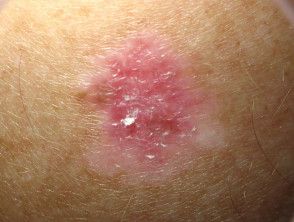
Reflectance Confocal Microscopy Enhances Detection Sensitivity for Flat Amelanotic/Hypomelanotic Melanoma Differential Diagnosis
Diagnostic accuracy of lesions was enhanced with combined dermoscopy and reflectance confocal microscopy, according to one study.
When combined with traditional dermoscopy, the use of adjunctive reflectance confocal microscopy is capable of increasing the overall diagnostic sensitivity of flat amelanotic and hypomelanotic melanoma (fAHM) differential diagnosis, according to a recent study. The study, published in the Journal of the European Academy of Dermatology and Venereology,1 shed light on a proposed model of identifying solitary flat pink lesions.
In 2020, researchers Lan et al conducted a systematic review and meta-analysis of scientific literature relating to the combined use of dermoscopy and reflectance confocal microscopy for fAHM detection.2 The review yielded a total of 7 studies and a total of 111 lesions. As a result of the review, researchers found that alone, both detection methods offer good diagnostic accuracy, high specificity, and moderate sensitivity, with reflectance confocal microscopy demonstrating more accuracy than dermoscopy in the diagnosis of these lesions.
However, the scientific literature to date has failed to define the diagnostic capabilities of these combined tools in the detection of fAHM.
In order to compare the diagnostic capabilities and accuracy of dermoscopy alone versus combined dermoscopy and reflectance confocal microscopy, researchers conducted a retrospective, single-centre, observational study. They aimed to propose an adjunctive model for the prediction of fAHM lesions within clinical practice settings.
The study was conducted at the Skin Cancer Center of the Arcispedale Santa Maria Nuova in Reggio Emilia, Italy. Researchers aimed to evaluate solitary, pink-colored macules or patches of the skin through various diagnostic techniques including clinical evaluation, dermoscopy, reflectance confocal microscopy, and histopathological examination. The study spanned from January 2011 to December 2022 and included consecutive patients meeting the predefined criteria.
Patients with suspected lesions observed during dermoscopy underwent real-time reflectance confocal microscopy evaluation, with excision programmed based on suspected diagnosis. Dermoscopic images were obtained utilizing standardized polarized dermoscopy, and reflectance confocal microscopy imaging was performed with the VivaScope 1500, with lesions assessed by 2 dermatologists.
The study included 184 patients with solitary flat pink lesions, with patient demographic data demonstrating a significant difference in average age between fAHM and non-fAHM patients. The most common diagnoses besides fAHM were basal cell carcinomas, dermatofibromas, and various other conditions.
A subgroup analysis of false-negative lesions following combined dermoscopy and reflectance confocal microscopy revealed misdiagnoses primarily as basal cell carcinomas. Specific diagnostic criteria observed through reflectance confocal microscopy included irregular honeycomb and disarranged epidermal patterns, roundish and dendritic pagetoid cells, and hyporeflective pagetoid cells.
Study limitations, as noted by study authors, include the study's retrospective design, the heterogeneous sample of equivocal solitary flat pink lesions, and the results' inability to be generalizable to all dermatological settings.
"Adjunctive RCM improves fAHM diagnostic accuracy among equivocal solitary flat pink lesions, with a higher sensitivity than dermoscopy alone: adjunctive RCM facilitates at least five extra melanoma diagnoses per 100 compared to dermoscopy alone," according to study authors Spadafora et al. "The proposed fAHM Index assists in improving diagnostic precision of solitary flat pink lesions."
References
- Spadafora M, Megna A, Lippolis N, et al. Dermoscopy and reflectance confocal microscopy of solitary flat pink lesions: A new combined score to diagnose amelanotic melanoma. J Eur Acad Dermatol Venereol. April 4, 2024. Accessed April 10, 2024. doi:10.1111/jdv.19991
- Lan J, Wen J, Cao S, Yin T, Jiang B, Lou Y, et al. The diagnostic accuracy of dermoscopy and reflectance confocal microscopy for amelanotic/hypomelanotic melanoma: a systematic review and meta-analysis. Br J Dermatol. 2020. Accessed April 10, 2024.
Recent Videos


Related Content