

|Articles|February 1, 2009
Post-transplant disorder a risk: Lymphoproliferative condition can be life-threatening
Author(s)Ilya Petrou, M.D.
Post-transplant lymphoproliferative disorder is lymphocyte proliferation in patients following post-transplant immunosuppression and is the most common neoplasm in pediatric organ transplant recipients. Due to the increased frequency of organ transplantation, dermatologists should be aware of the cutaneous presentation, as a timely diagnosis and treatment is crucial for a favorable prognosis.
Advertisement
Key Points
PTLD is essentially the result of immunosuppression in a transplant patient, and pediatric patients are especially susceptible to this type of B-cell lymphoma malignancy, most probably due to their exposure to certain infectious diseases.
"These patients may have never been exposed to the virus before, and then they receive a transplant from a donor who has EBV infection, passing on the virus to them," says Claudia Hernandez, M.D., assistant professor, department of dermatology, University of Illinois, Chicago.
Case study
Dr. Hernandez recently had a 14-month-old female OTR patient with liver and small bowel transplantation due to congenital gastroschisis and volvulus. Prior to procedure, the patient was seronegative for EBV, but the related living donor was EBV and CMV (cytomegalovirus) positive.
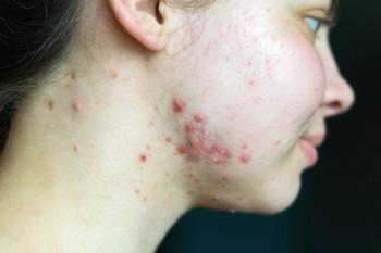
Approximately five months after transplantation, in the setting of immunosuppression therapy and rising EBV titers, the patient developed multiple erythematous, blanching nodules on the abdomen adjacent to her surgical scars.
Biopsy of a lesion showed a dense infiltrate of large cells in the dermal and subcutaneous layers characterized with prominent nucleoli, open chromatin and abundant cytoplasm, with mitotic figures being a common feature.
Immunohistochemical stains were positive for CD138, CD56, Ki67 and lambda chain restriction. Rare mature B cells (CD20) and rare T cells (CD3) were also present. The patient was thus diagnosed with a high-grade PTLD most consistent with plasmablastic lymphoma.
"Most OTR patients receive high doses of immunosuppressive agents to prevent organ rejection. One of the most important steps in PTLD treatment is to withdraw or reduce immunosuppressive therapy," Dr. Hernandez tells Dermatology Times.
In this case, the patient was initially suspected to have graft-versus-host disease and, thus, immunosuppression was increased. This increase allowed a tremendous rise in the EBV viral load from 449 copies/ml (upon admission) to 96,719 copies/ml, in spite of concomitant ganciclovir therapy.
The patient then received a course of rituximab as part of a children's oncology group protocol for PTLD due to her rapid clinical deterioration. Though these quick measures were taken, the patient developed multiple organ dysfunction syndrome and expired.
Only 48 hours had transpired between the presentation of skin lesions to the time of death.
First-line therapy
Most of the cases of PTLD are B-cell lymphomas. Rituximab is an anti-CD 20 monoclonal antibody therapy that specifically targets B cells. The first-line therapy in patients with PTLD is reduction of immunosuppression and the administration of antiviral agents. If that does not work, then one should try rituximab as a second line of therapy.
"A lot of transplant therapy is being performed nowadays and, therefore, we have to maintain a high index of suspicion when we see cutaneous manifestations such as those seen in this case," Dr. Hernandez says.
Risk factors
According to Dr. Hernandez, risk factors for developing PTLD include the type of transplant - with heart, lung (or combined) and intestinal transplantation ranking high - along with high levels of immunosuppression, increases in EBV viral load, and EBV seronegative recipient and EBV seropositive donor.
Disclosure: Dr. Hernandez reports no relevant financial disclosures.
Advertisement
Related Content
Advertisement


Latest CME


Advertisement
Advertisement
Trending on Dermatology Times
1
FDA Accepts Addition of Bemotrizinol as First New Sunscreen Ingredient in 20 Years
2
AI Analysis Reveals Menopausal Hair Loss Linked to Systemic Health and Inflammation
3
Dual-Action Microneedle Patch Shows Promise for Psoriasis in Preclinical Study
4
Novel Botanical Moisturizer Demonstrates Superior Efficacy Over Metronidazole in Phase 2 Rosacea Trial
5