

|Articles|June 20, 2022
New Insights into Pustular Psoriasis
Author(s)Nicholas Brownstone, MD
A new study aimed to evaluate the differences in the immunohistochemical staining (IHS) in patients with palmoplantar pustulosis and determine whether they can be divided into two distinct groups based on their histopathological profiles
Advertisement
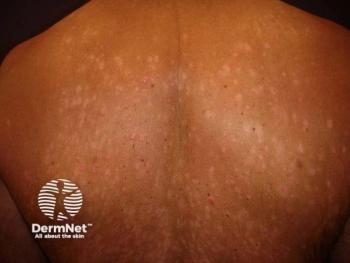
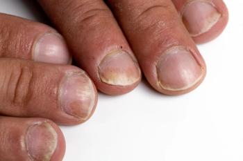
Many different forms of psoriasis exist including inverse/intertriginous psoriasis, scalp psoriasis, nail psoriasis, guttate psoriasis, erythrodermic and pustular psoriasis (which includes palmoplantar pustulosis and palmoplantar pustular psoriasis). All of these different forms of disease usually respond to the same treatment regimen which includes topical corticosteroids, oral therapies, biologic therapies and phototherapy. Psoriasis has many different causes and while there is evidence for a genetic link, in some cases its onset is idiopathic.1 Guttate psoriasis has even been shown to be trigged by infection.2
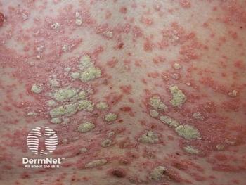
Given the varied presentations and etiologies of psoriasis, there is a theory that psoriasis is in fact many different but semi-related diseases. For example, while palmoplantar pustulosis and palmoplantar pustular psoriasis have similar characteristics, It is debatable whether palmoplantar pustulosis should be considered a distinct entity or should belong to the spectrum of pustular psoriasis.3 “Previously considered a subtype of psoriasis, important differences in the genetics and immune dysregulation between psoriasis and palmoplantar pustulosis have been identified, demonstrating that palmoplantar pustulosis may be a distinct entity. Further complicating the issues is the fact that the clinical phenotype of psoriasis on the palms and soles and palmoplantar pustulosis occurs along a spectrum with well demarcated plaques on one end, sterile pustules on the other and many patients with features of both”, said Megan Noe, MD, MPH, MS a Board-Certified Dermatologist and an Assistant Professor of Dermatology at Brigham and Woman’s Hospital/Harvard Medical School.
A new study in Annals of Dermatology aimed to evaluate the differences in the immunohistochemical staining (IHS) in patients with palmoplantar pustulosis and determine whether they can be divided into two distinct groups based on their histopathological profiles. Nineteen patients with palmoplantar pustulosis were enrolled in the study and a retrospective analysis was conducted using medical records and punch biopsy specimens. Inclusion criteria included patients with a diagnosis of palmoplantar pustulosis with neutrophil containing pustules. Patients were excluded if they did not have intact pustules or had another concurrent diagnosis (such as generalized pustular psoriasis). Skin samples were analyzed with seven antibodies: anti-α-3-nAChR, anti-IL-23, anti-IL-36R, anti-LL37, anti-IL-8, anti-CD3 and were also independently analyzed by two dermatologists for the degree of staining (which was classified on 4 point scale from negative to strong). Statistical analysis was conducted using principle component analysis (PCA), which is a multivariate statistical method that aims to consolidate high-dimensional variables into simpler variables named principle components (PC).
The authors found that the three main PCs accounted for 64% of the total variance in palmoplantar pustulosis. PC1 (pustular psoriasis properties) showed a higher correlation with IL-36R and PC2 (acrosyringeal/inflammatory properties) showed a higher correlation with α-3-nAChR, IL-8, LCN2, and CD3. PC3 (psoriasis properties) showed a higher correlation with IL-23. PC1 showed a statistically significant difference between the two groups (based on the Wilcoxon rank-sum test), with no significant differences in other PCs. Noe stated the following on these results, “this study highlights the complicated relationship between palmoplantar pustulosis and palmoplantar psoriasis, as the authors were not able to differentiate between the two diseases using a principal component analysis based on 7 IHC antibodies. They concluded that palmoplantar pustulosis and palmoplantar pustular psoriasis share the series of a inflammatory process.”
Interestingly, the results indicated that PC3 (the plaque psoriasis component) had a correlation with IL-23. Guselkumab, an anti-IL-23 monoclonal antibody, is FDA approved for the treatment of plaque psoriasis and has also shown to have efficacy in the treatment of palmoplantar pustulosis. A double-blind, randomized, placebo-controlled 24 week clinical trial of 49 patients with palmoplantar pustulosis showed that guselkumab resulted in significant improvements from baseline versus placebo at week 16.4 Moreover, IL-36R activation by IL-36γ can induce IL-23 cytokines, which plays a critical role in the pathophysiology of psoriasis.5 The authors found that PC1 (pustular psoriasis component) had a correlation with IL-36R and presented a significant difference between the two groups, which may represent a therapeutic target for pustular inflammatory diseases. Spesolimab, a humanized anti-interleukin-36 receptor monoclonal antibody is being studied for generalized pustular psoriasis.6 This medication may prove useful for palmoplantar pustulosis and palmoplantar pustular psoriasis but more studies are needed. “This overlap between psoriasis of the palms and soles (with and without pustules) and palmoplantar pustulosis creates a heterogenous and often unpredictable response to currently available psoriasis treatment in people with palmoplantar pustulosis”, added Noe. Therefore, more targeted and effective medications are needed for these disorders.
This study was not without limitations. Principally, PCA is strictly a statistical method and may not be indicative of the clinical reality. Furthermore, the interpretation of the PCs is mainly research dependent and the immunohistochemical analysis was only semi-quantitative. Finally, a larger patient population is necessary to better detect the immunological differences. Even though palmoplantar pustulosis could not be distinguished from palmoplantar pustular psoriasis, three meaningful PCs associated with the pathophysiology of palmoplantar pustulosis were identified. “Despite the similarities between the two diseases identified in this study, we know that that key differences also exist and additional research is necessary to better understand their relationship to one another”, Noe stated.
In conclusion, this study was unable to clearly distinguish palmoplantar pustulosis from palmoplantar pustular psoriasis, but the authors did identify three meaningful PCs. The PCs that were associated with the pathomechanism of palmoplantar pustulosis were PC1 (pustular psoriasis component), PC2 (acrosyringial/inflammatory component), and PC3 (plaque psoriasis component). To aid in the differentiation of palmoplantar pustulosis from palmoplantar pustular psoriasis in future studies, immunological studies with a larger patient population (compared to psoriasis) is a suggested by the authors. The ultimate goal of this study, and future ones, is to help better define the pathophysiology of pustular psoriasis to develop better therapies. Given that there are no current FDA approved medications for this condition, clinicians are forced to use off label medications with little data on safety and efficacy for pustular disease. While some of these therapies are fortunately effective, access may be an issue given that there is no FDA approval for their use in pustular psoriatic disease which stresses the need for more innovation in this area.
References
1. Dand N, Mahil SK, Capon F, Smith CH, Simpson MA, Barker JN. Psoriasis and Genetics. Acta Derm Venereol. 2020;100(3):adv00030. doi:10.2340/00015555-3384
2. Telfer NR, Chalmers RJ, Whale K, Colman G. The role of streptococcal infection in the initiation of guttate psoriasis. Arch Dermatol. 1992;128(1):39-42.
3. Navarini AA, Burden AD, Capon F, et al. European consensus statement on phenotypes of pustular psoriasis. J Eur Acad Dermatol Venereol. 2017;31(11):1792-1799. doi:10.1111/jdv.14386
4. Terui T, Kobayashi S, Okubo Y, Murakami M, Hirose K, Kubo H. Efficacy and Safety of Guselkumab, an Anti–interleukin 23 Monoclonal Antibody, for Palmoplantar Pustulosis. JAMA Dermatol. 2018;154(3):309-316. doi:10.1001/jamadermatol.2017.5937
5. Bridgewood C, Fearnley GW, Berekmeri A, et al. IL-36γ Is a Strong Inducer of IL-23 in Psoriatic Cells and Activates Angiogenesis. Front Immunol. 2018;9:200. doi:10.3389/fimmu.2018.00200
6. Bachelez H, Choon SE, Marrakchi S, et al. Trial of Spesolimab for Generalized Pustular Psoriasis. New England Journal of Medicine. 2021;385(26):2431-2440. doi:10.1056/NEJMoa2111563
Advertisement
Related Content
Advertisement

Latest CME


Advertisement
Advertisement
Trending on Dermatology Times
1
Study Suggests Non-Ablative Fractional Laser Reverses Epigenetic Signatures of Skin Aging
2
The Gut-Skin Axis: New Evidence Links Microbiota to Skin Disease via Inflammatory Cytokines
3
Bempikibart Shows Positive Phase 2a Results in Alopecia Areata
4
Social Media Mythbusters: PDRN Serums
5