New clinical syndromes
National report - Some of today's newest clinical syndromes highlight the fact that diseases of other organs, including the heart, often manifest on the skin, and these skin findings can offer clues to disease severity.
Paolo Romanelli, M.D., assistant professor of dermatology and cutaneous surgery, Miller School of Medicine, University of Miami, Miami, discusses six new syndromes: acquired brachial cutaneous dyschromatosis (ABCD), granular parakeratosis, hyperhomocysteinemia, calciphylaxis, nephrogenic fibrosing dermopathy and cardio-cutaneous mucinosis.
ABCD
"It is a condition that dermatologists usually see in middle-aged women as brown patches on their arms and forearms, which are not to be confused with melasma or sun damage. When they are asked, these patients will tell you that they have high blood pressure and are being treated with ACE inhibitors," Dr. Romanelli says.
Granular parakeratosis
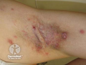
First described as granular parakeratosis in the axilla, the condition has since been identified in the groin and other parts of the body. Patients present with brown papules on the skin, which, when biopsied, reveal granular parakeratosis.
"Syndromes are usually defined by some basic defect, but, in this case, we do not yet know the cause," Dr. Romanelli says.
According to Dr. Romanelli, there are studies showing a defect from profilaggrin to filaggrin, which is similar to but different from the defect well-known for ichthyosis vulgaris.
"Granular parakeratosis was first described in 1991. It was described as a disorder of keratinization and is not a life-threatening problem, but rather usually a cosmetic concern," he says.
Hyperhomocysteinemia
Dr. Romanelli and colleagues at the University of Miami have witnessed the effects of hyperhomocysteinemia in cutaneous ulcers, which often results in resistance to standard ulcer treatments.
"Not too long ago, Dr. McCully described hyperhomocysteinemia as an important risk factor that can cause thrombosis and leg ulcers," he says.
The condition is treatable, according to Dr. Romanelli, with 800 mcg daily of folic acid.
"We had one patient whose homocysteine was extremely high, and this patient had ulcers on the fingertips. We treated the patient with folic acid and the condition improved," he says.
Calciphylaxis
Patients who have calcific uremic arteriolopathy (calciphylaxis), a serious condition that involves kidneys (resulting in renal failure), bone and skin, get large painful ulcers with eschar.
"When we do a skin biopsy, we see calcium deposits on the vessels on the dermis. These patients, unfortunately, almost invariably die," Dr. Romanelli says.
One of the new theories is that the culprit of calciphylaxis could be osteopontin, a protein that changes the wall of the vessels in the skin from muscular tissue to bone.
Nephrogenic fibrosing dermopathy
Described in 2000 at Yale University, New Haven, Conn., and the University of California, San Francisco, nephrogenic fibrosing dermopathy is a condition that affects patients who have renal failure and are undergoing dialysis or have had kidney transplants.
For some unknown reason, these patients develop induration and brown discoloration of the upper and lower extremities and sometimes of the trunk.
The wood-like texture of the skin on the upper and lower extremities can be severe and deforming. One of Dr. Romanelli's patients recently died from the condition.
Newsletter
Like what you’re reading? Subscribe to Dermatology Times for weekly updates on therapies, innovations, and real-world practice tips.


