

|Articles|June 1, 2017
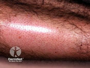
From mole to melanoma
There is a genetically intermediate state of melanocytic neoplasia and these findings could someday help doctors identify high-risk lesions to remove before they become malignant.
Advertisement
Benign nevi are clonal proliferations of partially transformed melanocytes, carrying only one or two of the multiple genetic alterations required to form melanoma. Melanomas arise from these precursors through the accumulation of additional mutations. The major carcinogen driving this accumulation is UV radiation, says Boris C. Bastian, M.D., professor of dermatology and pathology, University of California, San Francisco. Dr. Bastian presented study findings on the topic during the plenary session at the March 2017 annual meeting of the American Academy of Dermatology in Orlando, Fla.
Dr. Bastian and his colleagues also found evidence of an intermediate genetic state, in which lesions have more genetic alterations than seen in unequivocally benign lesions but fewer alterations than observed in malignant lesions. This intermediate state coincides with an intermediate histologic pattern, about which dermatopathologists tend to vary widely in their microscopic assessment.
“What this data shows, I think for the first time, is that there is a genetically intermediate state of melanocytic neoplasia. The concept that we go from white to black in melanoma does not seem to be correct. There is a step-wise evolution from a clearly benign lesion to a clearly malignant lesion, and there is a genetic state in between these two evolutionary stages,” Dr. Bastian says.
While further work is needed to precisely define the clinical and histopathological correlates of that intermediate state, these findings could someday help doctors identify high-risk lesions to remove before they become malignant.
Transition analysis
The subset of melanomas that arise from preneoplastic lesions typically originates from nevi or melanoma in situ. To analyze how a mole turns cancerous and how melanoma in situ becomes invasive melanoma, Dr. Bastian and colleagues selected melanomas with an adjacent nevus or melanoma in situ. They separately microdissected melanoma and precursor cells, and determined the mutation status of common cancer genes.
In a study published November 12, 2015 in the New England Journal of Medicine, Dr. Bastian and coauthors analyzed 37 of these hybrid tumors and compared genetic alterations in more than 200 areas.
Illustrating one exemplary case, he says: “This melanoma arose from a nevus that had only a BRAF V600E mutation, and during the progression to melanoma, it acquired a whole range of additional mutations. Most are silent mutations, but a few are additional pathogenic mutations, which disrupt additional signaling pathways.”
In the footprint of the genomes that the researchers sequenced, which is a little less than one-tenth of a percent of the entire genome, the researchers found about 10 mutations in the nevus. But the number of mutations significantly increased with the transition to melanoma.
“As we compared the benign to the malignant areas, the mutation burden increased. And when we look at the nature of the mutations, we see that most of the mutations are C to T transitions at dipyrimidine sites, which is the typical UV signature mutation,” he says. “So what we conclude from this analysis is that melanomas arise from these preneoplastic lesions through the accumulation of additional genetic alterations, and that UV radiation is the major mutagen that causes the additional genetic alterations.”
In essence, he says, nevi constitute clonal proliferations of partially transformed melanocytes, where a single melanocyte acquires a mutation of a dominant oncogene, which then leads to a limited clonal expansion. These initiating mutations affect the main signaling pathway controlling cell proliferation, which is the MAP kinase pathway and can occur at the level of the upstream receptor, at RAS or - most frequently - at BRAF.
Dr. Bastian emphasizes that the way melanomas evolve from pre-neoplastic lesions varies significantly among melanoma subtypes. During the meeting, showing a blue nevus-like melanoma that arose from a blue nevus, he said: “There are complete different mutations here. These lesions start with G protein mutations in GNAQ or GNA11, with subsequent progression through mutations in BAP1, SF3B1 or EIF1AX. This is an identical progression as in uveal melanoma, to which these neoplasms are closely related.”
In situ
The researchers also looked at melanomas that arise from melanoma in situ, rather than from nevi. While quite similar in the later phases, these lesions typically do not start with BRAF V600E. They have different mutations or BRAF or, more frequently, mutations in NRAS or loss of function mutations in NF1, he says.
Not all melanomas arise with a clearly discernable pre-neoplastic lesion, according to Dr. Bastian.
“There are lesions that rapidly emerge from skin that either historically or sometimes demonstrably has been uninvolved. It will be interesting to determine how these lesions evolve: if they arise from a field effect of transformed melanocytes or whether a subtle precursor is simply overrun by these lesions,” he says. “It’s important to note that melanomas of the skin are one of the most highly mutated cancers from the Cancer Genome Atlas project. The many mutations we find in these advanced melanomas could not have all [arisen] at once; they accumulate with time, and thus it is likely that even in melanomas arising without an apparent precursor, there is some yet-to-be-defined precursor state.”
The hope, according to Dr. Bastian, is that researchers can use this information to build a molecular taxonomy of melanoma, whereby they distinguish the various classes of melanoma and then delineate the different evolutionary trajectories within each separate class and identify specific genetic states associated with the progression from one state to the next. The result, he says, would be improved diagnostic accuracy and provide better ability to predict the likelihood that a lesion will metastasize. Â
Disclosure: Dr.Bastian reports no relevant disclosures.
Advertisement
Related Content
Advertisement


Latest CME


Advertisement
Advertisement
Trending on Dermatology Times
1
Social Media Mythbusters: Skin Cycling
2
Clinique's Daily Calm Merges Clinical Efficacy with Color Cosmetics for Sensitive Skin
3
What Dermatologists Need to Know About the First New Sunscreen Ingredient in Decades
4
First HA Injectable for Neck Wrinkles Cleared by FDA
5