
|Articles|October 10, 2016
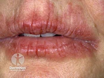
Melasma data strengthens therapeutic insight
Agents that can attack melasma on multiple fronts include retinoids, glucosamine, hydroquinone, topical steroids, phytosterol, glycyrrhetinic acid, niacinamide, soybean extracts, retinoids, salicylic acid and liquirtin, an expert says.
Advertisement
Addressing the multifaceted pathogenesis of melasma requires combining multiple treatment modalities, says Pearl E. Grimes, M.D., who spoke at MauiDerm this year.
"In 2016," Dr. Grimes says "we have come to realize that melasma is a far more complicated skin disease than we ever thought." She is director of the Vitiligo and Pigmentation Institute of Southern California and clinical professor of dermatology at The University of California Los Angeles David Geffen School of Medicine.
A decade ago, “We believed melasma was a condition that simply affected melanocytes, increasing the number of melanocytes, and melanin production, in affected areas. Now, most data - including mine - have shown that melanocytes are not increased. We simply have an increase in melanogenesis."1
Along with melanocytes, she says, key players driving the increased melanogenesis include keratinocytes, fibroblasts and even mast cells in the dermis. Additionally, "A host of genes are either up- or downregulated." These abnormalities can lead to increased expression of C-kit, also called stem cell receptor factor or CD 117. "If we look at genomic studies, we know that in addition to upregulation of pigment-cell genes, prostaglandin genes are upregulated. WNT pathway genes are also altered."
Conversely, she says, downregulation of lipid-related genes in affected skin compromises its barrier function. Most studies comparing biopsy samples of affected versus normal skin show increased sun damage in affected areas. Biopsies from affected skin also reveal increases in blood vessel markers (such as vascular endothelial growth factor/VEGF), dermal blood vessels and mast cells, she says.
Mast cells are extremely important in melasma because they release mediators ranging from histamine to the angiogenesis-driving factors VEGF, TNF-alpha and interleukin (IL)-8, says Dr. Grimes. "This is very new - the role that mast cells play in the pathogenesis of melasma."
Until about eight years ago, "We thought maybe the vascular component was related to products we were using on the skin. As we looked closer, we were seeing melasma in patients who had never been treated with these products."
Unfortunately, Dr. Grimes adds, "We don't have great tools to address melasma's vascular component. In Asia, topical and oral tranexemic acid are commonly prescribed and may impact the vascular component of melasma. In addition, vascular lasers such as the pulsed-dye laser (PDL) may also address the vascular component." She cautions that any visible or infrared light may spur angiogenesis. In general, she avoids aggressive laser treatments for melasma.
In recent years, Dr. Grimes says physicians have come to appreciate the role of visible light in the pathogenesis of hyperpigmentation. "Unfortunately we don't have highly effective visible light sunscreens," outside of iron oxide.
Far more fragile
Given the abnormal barrier function in patients with melasma, Dr. Grimes says, their skin is probably "far more fragile than we have ever realized. We've probably been too aggressive in treating this disease, particularly with lasers, light sources and deeper peels. Most evidence-based studies in melasma suggest that lasers and light sources should not be first-line therapy.2 However, there's a different paradigm in Asia," where laser treatments for melasma are extremely popular.
Specifically, she says, laser "toning" involves a 1,064 nm Q-switched laser operating at a low fluence, which requires many more sessions per patient than Western physicians typically perform. "The complication, according to recent studies, is the high incidence of leukoderma that's being induced by these treatments. It's a problem in Asia. We don't see nearly as much of it in the United States."
Most effective for melasma, say several large reviews, are therapies that combine various mechanisms of action, especially triple-combination bleaching (TCB; hydroquinone 4%, tretinoin 0.05%, fluocinolone 0.01%).3-5 A recent study further supports the high efficacy of triple combination bleaching, with no evidence of steroid-induced skin atrophy.6 More recently, an 18-patient study showed that combining TCB with PDL provided greater efficacy than TCB alone.7
Popular bleaching agents attack melasma on many fronts, Dr. Grimes says. For example, retinoids, glucosamine and hydroquinone target tyrosinase by downregulating its production, blocking its biological activity or inhibiting tyrosinase glycosylation, respectively. Topical steroids inhibit inflammation, as do phytosterol and glycyrrhetinic acid, she says. Niacinamide, soybean extracts and protease inhibitors block melanosome transfer, while retinoids, salicylic acid, lactic acid, glycolic acid and liquirtin increase epidermal turnover.
Along with increasing epidermal turnover and extrusion of pigment, Dr. Grimes says, chemical peels can provide synergistic adjuncts to topical regimens because chemical peels also may reduce inflammation.
Emerging ingredients for fighting melasma also offer multiple mechanisms, she says. Examples include tranexemic acid (anti-inflammatory, tyrosinase inhibition and degradation, plasmin inhibition) and cysteamine (tyrosinase and peroxidase inhibition). Other up-and-coming treatments for melasma include Lytera (SkinMedica), elure (lignin peroxidase, Syneron), Lumixyl (oligopeptide, Envy) and Perle Skin Brightening Cream (Neocutis), she says.
Establishing expectations
When counseling patients, "I emphasize that melasma is a chronic disease. That allows me to establish patient expectations. I emphasize, 'There is no cure. But don't despair - I can make every effort to stabilize it, treat it and make it better, and hopefully keep it in remission for a long time.' Patients are much more comfortable if you explain it that way."
She also prepares patients for the necessity of maintenance treatments during remission periods, followed by periodic flare-ups that require more active treatment.
"A clearing phase may take three to six months. That varies from patient to patient. But if I put a patient on a given therapeutic regimen, I have the patient return in four weeks. I typically like to see something happening" at this point. If a patient sees no results after three months of treatment, "I typically make a major adjustment in the patient's regimen."
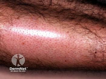
At the other end of the pigmentation spectrum, "Erythema dyschromicum perstans (EDP) is probably one of the most difficult - if not the most difficult - pigment condition that I treat. It's a lichenoid dermatitis that may be part of the lichen planus spectrum. Patients may develop it in response to some type of environmental allergen. My standard protocol is to patch test virtually every patient I see who has EDP." Treatments include topical and oral corticosteroids, lighteners, calcineurin inhibitors, dapsone, narrowband UVB phototherapy and clofazimine, she says.
Postinflammatory hyperpigmentation (PIH) differs greatly from melasma, Dr. Grimes says, because it's a response to inflammation produced by skin conditions such as acne. Often it's easier to treat than melasma because the pathogenic process has stopped. "If you treat the primary disease, you have a much better chance of treating and curing PIH."
Disclosure: Dr. Grimes has performed clinical research and/or served as a consultant for Allergan, Clinuvel, Galderma, Johnson & Johnson, Kythera, Procter & Gamble, Merz, Suneva and Valeant.
References
1.
2.
3. Rivas S, Pandya AG.
4.
5.
6.
7.
Advertisement
Related Content
Advertisement


Latest CME


Advertisement
Advertisement
Trending on Dermatology Times
1
Social Media Mythbusters: Skin Cycling
2
What Dermatologists Need to Know About the First New Sunscreen Ingredient in Decades
3
Multidisciplinary Care Anchors Treatment of Advanced cSCC
4
Clinique's Daily Calm Merges Clinical Efficacy with Color Cosmetics for Sensitive Skin
5