

|Articles|March 1, 2008
Melanoma: Seven-point checklist, genetic analysis are cornerstone of skin exam
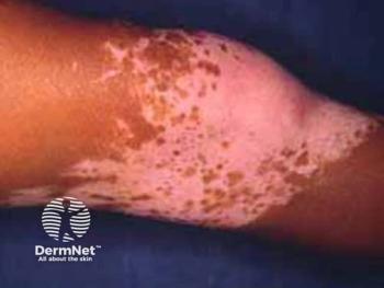
Melanomas can appear very different in the dermoscope and have different genetic mutations, depending on the amount of ultraviolet radiation insult received over the years.0
Advertisement
Key Points
According to one expert in the field, melanomas can show very different faces in the dermoscope examination, as well as in their genetic expression, depending on the anatomical location of the lesion.
"There are different types of melanomas in terms of genetic alteration, depending on where the lesions develop. This can be witnessed in their different dermoscopic patterns, reflecting a different pathogenesis," says Wilhelm Stolz, M.D., chief, department of dermatology at the München-Schwabing Hospital and professor of dermatology, University of Munich, Germany.
As Boris Bastian, M.D., and colleagues at the University of California, San Francisco, have shown, melanomas on skin without marked solar elastosis frequently show mutations in BRAF. These melanomas are predominantly located on the trunk and proximal extremities and occur in younger individuals.
By contrast, melanomas on chronically sun-damaged skin, such as the face and distal extremities of older individuals, as well as acral melanomas show a significantly lower frequency of BRAF mutations.
More recent analyses from that group have shown that 30 percent to 40 percent of the latter two categories of melanoma can have mutations or amplifications of c-KIT.
"These melanomas are not exclusive to these anatomic locations, but they are the most common for these specific areas, and most melanomas follow these rules and characteristics," Dr. Stolz tells Dermatology Times.
Dr. Stolz says the development of these three types of melanomas in these different anatomical locations is related to the different amount and intensity of sun exposure over the years, reflecting also a different genetic mutation in each type.
"If you have intermittent strong sun exposure or a number of sunburns over the years, you will see certain types of mutations, such as a BRAF mutation as seen in melanomas on the trunk.
"One can also see BRAF mutations in melanocytic nevi; however, the melanomas on the trunk exhibit a higher amounts of BRAF mutations," Dr. Stolz says.
Melanomas on the face can also have BRAF mutations, but the majority of the cases of the melanomas located on the face are slower-growing lesions, taking years to grow.
The role of sun exposure
According to Dr. Stolz, the sun exposure on the face is strong and may be considered stronger than sun exposure received on the trunk, but the difference is that it is a chronic sun exposure.
"Possibly, the immune defense mechanisms of the body and in the face are able to adapt to the long-term insults of the ultraviolet radiation. But the result is the development of another type of melanoma, different from those seen on the trunk and extremities, where there is chronically less cumulative ultraviolet radiation," Dr. Stolz says.
Lentigo maligna is a melanoma in situ and can be present for years before growing vertically into a nodular melanoma. In the past, lentigo maligna was, for the most part, only seen in very old individuals, but according to Dr. Stolz, this has now changed - possibly owing to changes in social behavior and sporting activities.
"Lentigo maligna and lentigo maligna melanoma represent a characteristic, histogenetic subclass of melanocytic malignancies.
"Lentigo maligna has a very similar and parallel relationship with malignant melanoma, very much as the relationship of actinic keratoses and squamous cell carcinoma.
"Lentigo maligna within the melanocytic system corresponds to the actinic keratosis within the squamous cell proliferation," Dr. Stolz says.
In melanomas on the trunk and extremities, the dermatologist should critically analyze the asymmetry of the lesion, the number of colors present and the number of different structural components.
Lentigo maligna - which commonly occurs on the face - has different dermoscopic features, including dark brown or black asymmetrically pigmented follicular openings, rhomboidal structures or an annular-granular pattern. Slate-gray dots and globules can also be seen, setting this apart from melanoma.
According to Dr. Stolz, if the dermatologist is prudent and patient in the dermoscopic analysis of such lesions, a much more accurate diagnosis can be made.
Newsletter
Like what you’re reading? Subscribe to Dermatology Times for weekly updates on therapies, innovations, and real-world practice tips.
Advertisement
Related Content
Advertisement


Latest CME


Advertisement
Advertisement
Trending on Dermatology Times
1
AbbVie Files for Vitiligo Indication, Putting Systemic Therapy Under Regulatory Review
2
Icotrokinra Shows Superior Efficacy Over Advanced Oral Therapies in New Psoriasis Meta-Analysis
3
Nutrafol Expands Portfolio with First and Only Hair Loss Supplement for Male Patients 50 and Older
4
Introducing Dermatology Times NP/PA Connect
5