MelaFind device raises concerns among some dermatologists, FDA
Although dermatologists have embraced mole-mapping techniques, including total body imaging and (to a lesser extent) dermoscopy, they appear to share the Food and Drug Administration's (FDA) concerns that non-dermatologists might misuse the MelaFind (MELA Sciences) device, which an FDA advisory panel recently recommended for approval in a split decision.
Key Points
Several physicians discussed the topic at the 2010 Cosmetic Surgery Forum in Las Vegas in December.
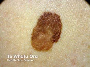
In diagnosing atypical nevi or changing moles, "We're interested in determining if a person has melanoma, because rapid treatment of melanoma is critical," says Cheryl Burgess, M.D., assistant clinical professor of dermatology, Georgetown University Hospital, and director, Center for Dermatology, Washington. In this regard, she says several technologies exist to help dermatologists decide whether a lesion requires biopsy or excision.
Total body imaging uses a single-lens reflex camera and imaging software to compare lesion changes over time to baseline photos, Dr. Burgess says.
Haines Ely, M.D., professor of dermatology, University of California, Davis, says, "I'm very much in favor of total body imaging." When a patient with dysplastic nevus syndrome or negative prognostic factors such as a family or personal history of melanoma presents with hundreds of moles, he says, "The melanomas aren't usually the moles they have. The melanomas usually develop de novo. And those machines are excellent at picking up a new spot. They can count all the spots and notice a change."
One such machine, the Melanoscan (Melanoscan), takes serial whole-body photographs over time. Dr. Ely says that with this method, "New lesions or lesions which have changed can be spotted quickly, and it is a true skin cancer screening device. In conjunction with a survey which uses clinical judgment, personal history of skin cancer, first-degree relatives with melanoma and total number of moles, the Melanoscan can provide a true risk assessment for development of melanoma. It has been shown to find melanomas when they are very thin or in situ (Drugge RJ, Nguyen C, Drugge ED, et al. Dermatol Online J. 2009;15(6):1)."
Magnification techniques
Dermoscopy uses magnification to increase diagnostic accuracy, Dr. Burgess says. In particular, she explains, dermatoscopes typically use LED light, occasionally with oil applied to the mole to enhance its features. This technology can highlight features such as the blue-white veil of melanoma or abnormal blood vessel development that tends to occur in pre-melanoma lesions, she says.
High-resolution ultrasound and confocal scanning laser microscopy also can help increase physicians' diagnostic accuracy, Dr. Burgess says. However, she notes, it's rare for dermatologists with small private practices to own such equipment, which is generally only available at larger teaching and research institutions.
MelaFind debate
Another diagnostic tool for melanoma, MelaFind has met with controversy, Drs. Burgess and Ely say. According to its manufacturer, this device captures, displays and stores multispectral and reconstructed RGB digital images of atypical pigmented skin lesions. Using automatic image analysis and statistical pattern recognition, the device was designed to differentiate between "positive" lesions - cutaneous malignant melanoma and high-grade dysplastic nevi (as well as atypical melanocytic proliferation and hyperplasia) - and less worrisome pigmented lesions, its manufacturer says.
Other computerized tools for recognizing abnormal pigmented lesions exist, Dr. Burgess says, "But the MelaFind is a little different. In simple terms, it provides a mini-MRI of the mole. It can scan images of an intact nevus within the skin.
"Overall, when tested against dermatologists, MelaFind performed better; however, the device missed a biopsy-confirmed diagnosis of melanoma (Monheit G, Cognetta AB, Ferris L, et al. Arch Dermatol. 2010 Oct 18. [Epub ahead of print])," Dr. Burgess says. In the product's pivotal trials, it demonstrated 98.3 percent biopsy sensitivity to melanoma and high-grade lesions, with a biopsy specificity of 10.8 percent, versus 5.6 percent for dermatologists.
Determining dermatologists' biopsy sensitivity was impossible in the product's pivotal trial, but in a companion reader study, this figure was 72 percent, versus 97 percent for MelaFind (p <0.0001), according to the MelaFind package insert.
Newsletter
Like what you’re reading? Subscribe to Dermatology Times for weekly updates on therapies, innovations, and real-world practice tips.


