
|Articles|June 1, 2013
Lentigo maligna removed with wider surgical margins
Subungual melanoma can be managed without the amputation of the distal digit, according to an expert who spoke at the 7th annual Canadian Melanoma Conference.
Advertisement
Banff, Alberta - Subungual melanoma can be managed without the amputation of the distal digit, according to an expert who spoke at the 7th annual Canadian Melanoma Conference.
The European experience and some experience at centers in the United States reveals subungual melanoma (which represents about 0.7 to 3.5 percent of melanoma subtypes) can be managed with en bloc resection of the nail instead of partial amputation, says Thomas G. Salopek, M.D., F.R.C.P.C., professor in the division of dermatology and cutaneous sciences, University of Alberta, and director of the Melanoma Clinic, University of Alberta, Edmonton.
More specifically, wide excision of the entire nail unit can be performed, with a safety margin of 5 mm to 10 mm, without the need for bone resection. A full-thickness skin graft is taken from the arm. No recurrences have occurred with this technique in seven cases with a mean follow-up time of 45 months (Sureda N, Phan A, Poulalhon N, et al. Br J Dermatol. 2011;165(4):852-858).
“It preserves function,” says Dr. Salopek, noting preservation of function would be significant for some professionals like musicians.”
Despite the absence of randomized, controlled trials to validate the appropriate management of subungual melanoma, standard treatment in North America has been partial amputation of the digit, explains Dr. Salopek, adding that an additional 62 cases in the medical literature demonstrate nonamputative management of subungual melanoma is safe and functionally superior to amputation.
Centers in Europe and some in the United States are currently offering nonamputative, conservative management of in situ or invasive subungual melanoma less than 1 mm in thickness.
In addition to function, patients and physicians have found the alternative approach to partial amputation produces a superior cosmetic result. “Cosmetically, it looks better as well,” Dr. Salopek says. “Most of the finger is left intact.”
Dr. Salopek emphasizes, however, the approach is designed for thin melanomas, those that are less than 1 mm, and is not meant for melanomas that are very deep.
“If we find the tumor is more aggressive on wide excision then we go back to the basic (management) principles,” Dr. Salopek says. He notes the approach is a suggestion for clinicians, not an official guideline.
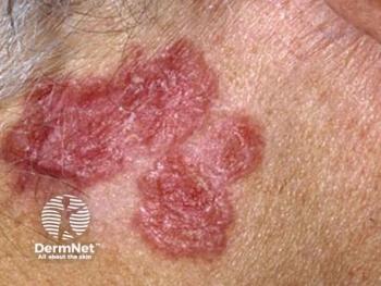
Dr. Salopek also discussed the ideal management of lentigo maligna, an area that is controversial. Lentigo maligna typically affects skin that is actinically damaged, he says. Despite generous wide local excision, atypical melanocytes are often noted near the surgical margins. Re-excision specimens show similar results.
The current recommendation is wide-local excision, with a 5 mm margin, but this practice is not evidence-based, Dr. Salopek suggests. Indeed, the practice is based on a consensus panel from the 1990s.
“There is no scientific basis for this,” Dr. Salopek says. “It is entirely based on opinion. There are no trials to validate or confirm the appropriateness of 5 mm margins.”
The results of a retrospective investigation of 1,072 patients found that standard surgical excision of melanoma in situ with 9 mm margins of normal-appearing skin would result in the complete removal of 99 percent of melanomas in situ. In contrast, excision with 6 mm margins would clear only 86 percent of melanomas in situ (Kunishige JH, Brodland DG, Zitelli JA. J Am Acad Dermatol. 2012;66(3):438-444).
An alternative approach to managing lentigno maligna in situ is called staged margin-controlled excision, which is also known as the “spaghetti technique.” This technique allows for 100 percent margin assessment.
The technique is reserved primarily for the face, a site where the cosmetic result is more of a priority to patients than sites on the body, Dr. Salopek says.
“You want to make sure you get all of the tumor, but you don't want to be excessive,” he says. “You don't want to be removing too much tissue. In contrast, if it is on the trunk, you don’t need to worry about a few millimeters.”
Although Mohs’ micrographic surgery is a reasonable alternative for treating lentigo maligna, with recurrence rates being quite low, many surgeons who perform Mohs’ surgery are not comfortable performing this surgery for melanoma (Temple C, Arlette JP. J Surg Oncol. 2006;94(4):287-292).
Disclosures: Dr. Salopek reports no relevant financial interests.
Advertisement
Related Content
Advertisement


Latest CME


Advertisement
Advertisement
Trending on Dermatology Times
1
KT-621 in Atopic Dermatitis: What Early EASI and Pruritus Data Show
2
What Dermatologists Need to Know About the First New Sunscreen Ingredient in Decades
3
Filling the HS Treatment Gap: Ruxolitinib Targets Early-Stage Disease
4
Once-Daily Zasocitinib Rivals Injectable Biologics for Skin Clearance, Phase 3 Data Show
5