
|Articles|February 1, 2014
Laser, light treatment hasten resolution of purpura
Purpura, whether after trauma or an aesthetic procedure, is undesirable to patients. The ecchymoses can last for up to two weeks, and dyspigmentation can persist even longer.
Advertisement
Joely Kaufman, M.D.
Purpura, whether after trauma or an aesthetic procedure, is undesirable to patients. The ecchymoses can last for up to two weeks, and dyspigmentation can persist even longer.
Jeremy Green, M.D.
Fortunately, there are laser and light options that utilize the principle of selective photothermolysis to target hemoglobin within extravasated red blood cells and speed the resolution of purpura. Despite their widespread use, however, there is a paucity of published literature on the topic.
Kevin Smith, M.D.
The earliest publication is from DeFatta and colleagues who reported their experience with the 595 nm yellow light pulsed dye laser (PDL) to treat the ecchymoses of 20 patients following facial plastic surgery (DeFatta RJ, Krishna Srinivasan, Williams EF. Arch Facial Plast Surg. 2009;11(2):99-103). At postoperative day (POD) five or six, the bruise was divided and part was treated with a PDL (V-beam, Candela) at 6 J/cm2, 10 mm spot, 10 ms pulse duration, and cryogen 30 milliseconds with a 20 millisecond delay for three passes.
The patients returned 48 to 72 hours later for PDL of the untreated areas at the same parameters, and had a final follow-up 48 hours later. Three independent blinded observers graded photographs obtained using a purpura scale created by the investigators. There was a statistically significant 63 percent reduction in ecchymosis score, and it was found that the PDL performed earlier (POD five) was more efficacious. Minimal edema but no dyspigmentation was noted.
In their discussion the authors mentioned an unpublished pilot study where they treated the purpura as it started to appear on POD two or three. They found that the ecchymosis continued to worsen over the next two to three days, so they defined POD five as the time of initial treatment for this study. The authors theorized that the deeper dissection plane of facial plastic surgery combined with the postoperative tissue edema rendered the PDL less effective until swelling had reduced and the extravasated erythrocytes migrated more superficially and were lysed.
Treating ecchymoses
Geronemus and colleagues published their experience with the same PDL in 10 patients who had ecchymoses from traumatic injury or a cosmetic procedure (Karen JK. Dermatol Surg. 2010). Six patients were treated 48 hours after the initial insult and the remaining four were treated at 72 hours at parameters more robust than the previous study: 7.5 J/cm2, 10 mm spot, 6 ms pulse duration, and cryogen 30 milliseconds with a 20 millisecond delay for a single pass. They followed the patients 24, 48 hours and seven days after the procedure, and found that accelerated bruise resolution was evident as soon as six hours after laser treatment.
Utilizing a scale created by the investigators, they found at 24 hours the average improvement was 62 percent in treated and 13 percent in untreated bruises. At 48 hours the average improvement was 76 and 36 percent, respectively. Two patients experienced minor transient crusting.
In their discussion the authors noted that the yellow color change in bruises is due to the conversion of hemoglobin (one absorption peak near 595 nm) to bilirubin (broad absorption peak at 460 nm). For this reason they believe earlier violaceous to erythematous bruises respond better to PDL. They also theorize that the bruises due to minor trauma or nonsurgical cosmetic procedures (i.e. injectables) are shallower and are accompanied by less edema than those induced by facial plastic surgery. Therefore the laser energy is more readily absorbed by the target chromophore and these ecchymoses are more amenable to earlier PDL treatment.
The most recent report on lasers for bruise reduction comes from a group at Baylor University (Mayo TT, Khan F, Hunt C, et al. Dermatol Surg. 2013;39(10):1459-1464). The investigators used the same PDL as the previous two studies to induce purpura at 6.5 J/cm2, 7 mm spot, 0.45 ms pulse duration cooling 30 ms/20 ms in 6, 2 x 2 cm zones on the lower abdomens of 17 patients. Each bruise was randomly treated with a cold compress, hydrogen peroxide soaked gauze, or a bruise serum for 10 minutes immediately after the bruise was induced or with a PDL (6.5 J/cm2, 7 mm spot, 6 ms, 30/20) 30 minutes after purpura induction.
Two blinded evaluators graded the bruises at 30 minutes, three days and seven days. There was no significant reduction in bruise duration versus control in the three other interventions, whereas the authors actually found a statistically significant increase in bruise time to resolution in the PDL treated group. The authors hypothesized that the application of PDL light energy so shortly after bruise induction may actually create an additive effect by disrupting hemostasis.
Evaluating types of light
It should be noted that visible light lasers and intense pulsed light devices with cut-off filters that cause the majority of light to be emitted in the visible light spectrum are safest when used in patients with Fitzpatrick skin types I to III. They can be used with caution in type IV, as melanin is a competing chromophore for short wavelength visible light.
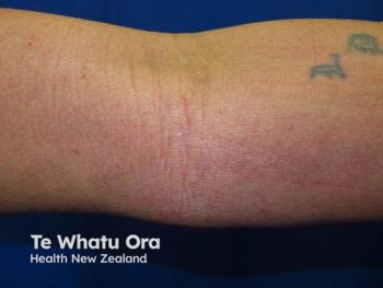
The authors of this column prefer to treat post-procedural and traumatic purpura with a 532 nm green light potassium titanyl phosphate (KTP) laser (Excel V, Cutera Lasers). One author (KCS) recently treated a five-day-old traumatic ecchymosis of the popliteal fossa with three different types of light to further understand optimal treatment.
The bruise (Figure 1) was divided into quadrants and treated with KTP laser at 14 J/cm2, 9 mm spot, 20 ms, 1064 nm neodymium yttrium aluminum garnet (Nd:YAG) laser at 60 J/cm2, 9 mm spot, 40 ms, intense pulsed light (IPL) with a short-wavelength cut-off filter at 20 J/cm2, 10 x 13 mm spot, program A (LimeLight, Cutera Lasers), versus an untreated control.
The following day (Figure 2), there was some minor improvement in the IPL- and Nd:YAG-treated sections, but considerable improvement in the KTP treated area. At that time all quadrants were treated with the KTP laser at the same settings, and only one hour later there was dramatic resolution of the treated areas (Figure 3).
The other authors (Dr. Green and Dr. Kaufman) sought to further ascertain optimal KTP parameters by treating a three-day-old traumatic bruise (Figure 4) with one half at the previously mentioned KTP settings, and the other half at the settings we had been using for bruises, 7 J/cm2, 10 mm spot, 10 ms. There was slightly more procedural discomfort with the higher fluence, longer pulse duration setting. The following day both sides improved from the treatment but the higher fluence setting was more impressive (Figure 5). The authors look forward to additional investigations that study the optimum device, parameters and timing of laser/light treatment of purpura after initial insult to further enhance our abilities to hasten the resolution of this undesirable sequela.
Disclosures: Drs. Kaufman and Green have performed research and served on the speakers’ bureau for Cutera Lasers.
Advertisement
Related Content
Advertisement

Latest CME


Advertisement
Advertisement
Trending on Dermatology Times
1
Chronic Hand Eczema: Treatment Gaps, New Options, and Clinical Trial End Points
2
Mona Shahriari, MD, FAAD: Matching the Right AD Therapy to the Right Patient
3
The Side Effect of Doing Nothing: Reframing Atopic Dermatitis as a Systemic Disease
4
First HA Injectable for Neck Wrinkles Cleared by FDA
5