
|Articles|April 27, 2015
Homing in on Targeted Gene Therapy for Congenital Melanocytic Nevi
New research data sheds light on potential gene therapy for congenital melanocytic nevi.
Advertisement
Although NRAS mutations are frequently found in congenital melanocytic nevi, new research data has revealed that BRAF mutations are also associated with the development of these nevi as well as with neurocutaneous melanocytosis. According to one expert in the field, these breakthrough findings could potentially open the door in the future for BRAF targeted therapies in select congenital melanocytic nevi and neurocutaneous melanocytosis cases that test positive for the BRAF mutated gene, offering much needed hope for this patient population.
Familiarize yourself with CMN and NCM
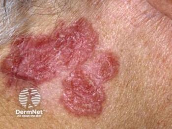
Giant Congenital Melanocytic Nevus (GCMN) with extensive nodularity involving the upper back and neck.
“Even though dermatologists are somewhat familiar with CMN and NCM, the average clinician may be overwhelmed when confronted with such a patient regarding therapy and management. Therefore, it is important that clinicians not only recognize this significant issue but also become more familiar with the current treatment approaches as well as those in the pipeline,” said Miguel Reyes-Múgica, M.D., Chief of Pathology and Director of Laboratories, Children's Hospital of Pittsburgh of UPMC, Pittsburgh, Penn.
Congenital melanocytic nevi (CMN) are benign proliferations of mutant melanocytes (nevomelanocytes) that are typically present at birth or develop shortly after birth, and can loosely be categorized into small (<1.5cm diameter), medium (1.5-10cm), large (11-20cm), and giant (>20cm diameter) lesions.
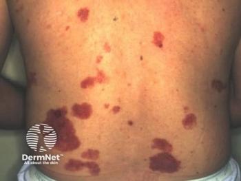
GCMN with multiple nodules involving the lower back and gluteal area.
Large and giant CMN lesions are particularly associated with a set of complications that include decreased sweating, xerosis, pruritus, and skin fragility. Patients also experience altered or diminished tissue growth due to the hamartomatous and infiltrative nature of the lesions; a higher complexity of surgical removal; and childhood psychological problems secondary to cosmetic issues and social stigmatization.
Congenital melanocytic nevi are also associated with an increased lifetime risk of malignant transformation to melanoma as well as the risk for neurocutaneous melanocytosis (NCM), a proliferation of nevomelanocytes in the leptomeninges and brain parenchyma, further underscoring the urgency to address these lesions appropriately and in a timely manner.
High power view of a temporal lobe from a patient with NCM showing pigmented cells infiltrating the brain parenchyma. Note melanin pigment also present within neurons (H&E, 40X).
“The biggest worry with CMN, although it may not be the most important one or the most frequent problem you see, is malignant transformation of the nevi. However, another significant concern is the stigmatization of these patients, as well as the limited treatment options of these lesions, which can be difficult to manage because surgeries can be quite challenging to perform and are typically multiple,” Dr. Reyes-Múgica said.
Individualized Treatment
Hairy GCMN involving the entire back. Satellites are evident in the extremities.
According to Dr. Reyes-Múgica, these typically complex and staged surgeries can also sometimes be disfiguring. The success of treatment often depends on the location of the nevus and its unique pattern or thickness, as well as the individual patient’s idiosyncratic ability to repair tissues.
“There are several different factors that need to be taken into consideration when deciding on the appropriate treatment approach for CMN lesions. Each case must be approached individually as no one treatment strategy fits all,” Dr. Reyes-Múgica said.
Dr. Reyes-Múgica and colleagues recently published their results of a prospective study that investigated the association between the standardized clinical features of CMN and large/giant CMN in a large patient cohort, with the mutational status of NRAS Q61 and BRAF V600 in nevi lesions.1
The ongoing study included 66 patients (40 female and 26 male) with 8 medium, 21 large, and 36 giant CMN lesions. One patient had only a melanocytic lesion from the brain and no clinical data. The main nevus size ranged from 6-80cm (mean 31.5cm), and satellite nevi count ranging from 0-300 (mean 58.9). At the time of initial surgery, patient ages ranged from 6 months to 18 years, with 51 patients younger than 3 years of age. Patient demographics included 48 Caucasians and 15 Asians, with information about race unavailable in 3 patients.
Classic low power histological view of a GCMN. Note the impressive thickness of the lesion, involving subcutaneous tissue and replacing dermal components (H&E, 4X).
Data showed that NRAS and BRAF mutations were present in 51 (77.3%) and 5 cases (7.6%), respectively, with 10 cases (15.2%) negative for both mutations. While NRAS mutations were found in 5 (62.2%) of 8 medium, 15 (76.2%) of 21 large, and 29 (80.6%) of 36 giant CMN lesions, BRAF mutations were seen in 1 (4.8%) of 21 large, and 4 (11.1%) of 36 giant CMN, with no BRAF mutations found in medium CMN lesions. While 100% of the BRAF mutations were found to be associated with increased dermal/subcutaneous nodules, only 32% of the NRAS-mutated nevi were associated with this finding. Results also showed that NCM affected 16 (24.2%) of 66 patients, where the NRAS and BRAF mutations occurred in 12 (75%) and 2 (12.5%) patients, respectively, with 2 patients (12.5%) negative for both mutations.
Proliferative nodule arising in a GCMN. Note the upper portion with scattered nevus cells surrounding a hair follicle in the upper center. The lower portion of the image shows a highly cellular lesion with small nevus cells (proliferative nodule) (H&E, 10X).
These results are important because not only could an association between BRAF mutations and large/giant CMN be identified, but there is also a clear association with the BRAF-mutated gene and NCM. Although the proportion may still be predominantly NRAS-mutated, Dr. Reyes-Múgica said that the BRAF-mutated nevi could potentially be approached differently with potential targeted treatment, such as BRAF inhibitors that could ultimately either prevent or decrease the proliferation of nevus cells.
GCMN stained with HMB45 immunohistochemistry. The lesion involves fascial plane and replaces the subcutaneous tissue and dermis (HMB45, 4X).
Brain cortex of a patient with neurocutaneous melanocytosis (NCM) showing pigmented NCM cells in the leptomeninges and infiltrating through the perivascular Virchow-Robin spaces (H&E, 4X).
Disclosures:
Dr. Reyes received a donation (20K) from Mr. and Mrs. Travis Bailey, which was matched by Nevus Outreach, Inc., to support his Tissue bank (a total of 40K). His work was partly supported by that donation.
References:
1. Salgado CM, Basu D, Nikiforova M, et al. BRAF mutations are also associated with neurocutaneous melanocytosis and large/giant congenital melanocytic nevi. Pediatr Dev Pathol. 2015;18(1):1-9.
Advertisement
Related Content
Advertisement


Latest CME


Advertisement
Advertisement
Trending on Dermatology Times
1
What Dermatologists Need to Know About the First New Sunscreen Ingredient in Decades
2
Social Media Mythbusters: Skin Cycling
3
The Expanding Role of Immunotherapies for Non-Melanoma Skin Cancers
4
Stapokibart Successfully Treats Refractory CSU After Omalizumab Failure in New Case Report
5