

|Articles|October 2, 2021
Grand Rounds in Dermatology: Maui Derm NP+PA Fall 2021
Author(s)Katie Hobbins
In a grand rounds presentation at the Maui Derm NP+PA Fall 2021 conference, Seemal Desai, MD, FAAD, Raegan Hunt, MD, PhD, Ted Rosen, MD, and Matthew J. Zirwas, MD, discussed some of their challenging cases.
Advertisement
In the presentation “Grand Rounds in Dermatology” at the Maui Derm NP+PA Fall 2021 conference, Seemal Desai, MD, FAAD, clinical assistant professor in the Department of Dermatology at the University of Texas Southwestern Medical Center, Dallas, Texas; Raegan Hunt, MD, PhD, chief of service, pediatric dermatology, Texas Children’s Hospital, and Baylor College of Medicine, Houston, Texas; Ted Rosen, MD, professor of dermatology, Baylor College of Medicine, Houston, Texas; and Matthew J. Zirwas, MD, director, Ohio Contact Dermatitis Center, and a member of the North American Contact Dermatitis Group, brought forward some of their challenging cases.1
Rosen highlighted 2 cases of Black patients presenting with white spots on the face and areas of depigmentation.
The first patient was a 37-year-old Black woman with a 4-month history of a slightly itchy rash. Medical history showed that she wasn’t currently on any medication and was in good health. She had also previously gotten an atrial septal defect repaired and has an anxiety disorder.
The second patient was a 73-year-old Black man with a 2-year history of a slightly itchy rash, and was not currently on my medication. However, he previously had prostate cancer, mild type 2 diabetes controlled by diet, and hypertriglyceridemia.
Rosen ordered a biopsy for both patients.
Patient 1’s biopsy showed unremarkable epidermis. The dermis shows “naked” granulomas, which Rosen explained is classic for a diagnosis of sarcoidosis.
Patient 2’s biopsy showed orthohyerkeratosis, and elongated rete ridges. The dermis contained sparse lymphocytic infiltrate, clinically warranted lichen simplex chronicus, and no decreased melanocytes. Due to these findings, Rosen diagnosed the patient with post-inflammatory dyschromia.
Zirwas put forth a case involving a 72-year-old female patient presenting with over 30 years of hand dermatitis. Starting in her late 30s while working as a nurse, he said the issues remained mostly the same over time and relief was only seen after using high dose oral steroids. However, when the steroid dose is reduced to below 30 mg per day, the dermatitis recurs.
She is currently under clobetasol under inclusion, had failed tacrolimus under occlusion, and had negative comprehensive patch testing.
There was no effect when prescribed methotrexate up to 15 mg per week. Additionally, she was treated with thalidomide to no effect, but she did develop peripheral neuropathy.
Zirwas prescribed mycophenolate 1500 mg over 3 months to no effect. He noted that since the patient weighed only 50 kg, 1500 mg is considered a high dosage and increasing it to 2000 mg would unlikely be more effective.
Next, he prescribed modified cyclosporine at 100 mg. The patient saw a 40% decrease in hand dermatitis, however, creatinine doubled. When cutting the dose in half, the creatinine normalized, but the hands returned to baseline.
Zirwas said that after all these treatments, he reflected on which questions he had not yet asked: whether she, or anyone else in her family have any allergies or asthma. The answer was yes.
Based on this revelation, he came to a diagnosis of atopic dermatitis, despite novel disease presentation.
He administered a shot of Kenalog and started her on dupilumab.
Hunt highlighted the case of a 11-year-old boy with a 6-week history of intermittent “prickling” rash, with additional symptoms including: fever, fatigue, anxiety, weight loss, photophobia, oral ulcers, chest pain, vomiting, diarrhea, myalgias, and arthralgias.
There was no reported autoimmune disease in his family’s medical history and was not on any medications.
A physical exam showed coalescing, annular, erythematous, and dusky patches. He had bilateral conjunctival injection, was tachypneic, and anxious.
She noted that the child had previous accidental mercury exposure.
Hunt ordered a biopsy, which found perivascular neutrophilic infiltrate with vascular fibrin deposition consistent with leukocytoclastic vasculitis (LCV).
Laboratory findings and imagining showed higher blood and urine mercury levels (72 ug/L, normal <=10.0ug/L and >80 ug/L, normal <=5.0 ug/L, respectably), an ANA of 1:1280, leukopenia, thrombocytopenia, transaminitis, Coombs positive hemolytic anemia, anti-phospholipid antibodies, proteinuria, bilateral pleural effusions, and pericardial effusion, low C3/C4, and low C1q.
Due to these findings, Hunt diagnosed the patient with hypocomplementemia Urticarial Vasculitis (HUV).
HUV is associated with renal, pulmonary, musculoskeletal, and gastrointestinal (GI) involvement. Hunt explained that mercury exposure can induce or exacerbate systemic autoimmune disease or lupus-like syndrome in genetically susceptible strains of mice. However, she cautioned, no prior descriptions of this phenomenon in humans have been reported in the literature.
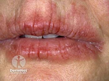
One case that Desai highlighted involved a woman in her mid-40s with chronic hair loss with brown and purple hyperpigmentation of the upper forehead. He presented that the hair loss—most likely alopecia areata— wasn’t as much of a concern as the pigmentary changes.
“What I want you to be aware of is when you start to see pigmentary issues in patients of color, look for that purple hue, that purple is really what's going to give you the clue on active inflammation because in most of these patients, you're not going to see redness like you would in a lighter skin tone individual such as erythema,” Desai said. “You tend to see more purple, and that purple is where you need to treat and also where you probably need to biopsy because that's where the active inflammation is.”
The pigmentation traveled down past her head, onto the spine of the back and front of the abdomen and stomach areas.
“What you're dealing with here is really a diffuse pigmentary anomaly with hyperpigmented, brown to violaceous and some graphite gray colored areas,” he explained.
He diagnosed the patient with lichen planus pigmentosus, not to be confused with lichen planus.
“Lichen planus is where you get the itchy purple red bumps on the wrists in the trunk,” Desai said. “[It’s] is not the same thing as lichen planus pigmentosa,” Desai said. “Let's say you have someone who comes in with classic lichen planus itchy papules. You treat them, they get better, and it leaves behind some dark marks. Fine that happens all the time. That is called lichen planus resolving with post inflammatory hyperpigmentation. that is not like lichen planus pigmentosus and I see that happening a lot when I get referrals.”
To treat this, he explained his go-to regimen includes isotretinoin 20 mg per day over 6 months. He also advises patients to wear sunscreen and antioxidants, and application of topical lightening agents and topical steroids.
Reference:
1. Desai S. Hunt R. Rosen T. Zirwas M. Grand Rounds in Dermatology. Session presented at: Maui Derm NP+PA Fall 2021 conference Program; October 1, 2021; Accessed October 1, 2021. Asheville, North Carolina
Advertisement
Related Content
Advertisement


Latest CME


Advertisement
Advertisement
Trending on Dermatology Times
1
Social Media Mythbusters: Skin Cycling
2
First HA Injectable for Neck Wrinkles Cleared by FDA
3
Clinique's Daily Calm Merges Clinical Efficacy with Color Cosmetics for Sensitive Skin
4
Oral STAT6 Degrader KT-621 Delivers Biologics-Like Efficacy in Moderate to Severe AD
5