Fungal Infections: Current therapies, solutions
San Diego - A multitude of fungi live in our ecosystem, and we can hope for peaceful coexistence with them - staying free from infection - but this is not always the rule. Sometimes when the immune system takes a turn for the worse fungi seize the opportunity to multiply and clinical presentation is the result.
At the American Academy of Dermatology's Summer Academy '06, Mahmoud A. Ghannoum, Ph.D., E.M.B.A., spoke in detail on the superficial, deep and systemic fungi that clinically affect patients, the current therapies available to the dermatologist and how they are employed to successfully combat these infections. He is a professor in the department of dermatology, director of the Center for Medical Mycology and co-director of the Skin Diseases Research Center at Case Western Reserve University in Cleveland.
According to Dr. Ghannoum, fungal infections are classified into superficial, subcutaneous, opportunistic and systemic. It is the superficial fungi that are of particular interest to the practicing clinician, because they are the most rampant and the most common reason why a patient with a fungal infection may seek out a dermatologist for help.
Causes, symptoms
Tinea barbae is a superficial infection of the bearded area of men and is caused by Trichophyton verrucosum and Trichophyton mentagrophytes.
The infection presents clinically as erythematous patches of the face and neck, showing scaling, fragile lusterless hairs and a tendency to folliculitis.
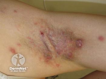
Tinea corporis is most commonly caused by Trichophyton rubrum, Tricophyton mentagrophytes and Epidermophyton floccosum, with a cutaneous presentation varying from scattered follicular to well-defined annular or doughnut-shaped lesions. Tinea cruris is also caused by Trichophyton rubrum, Tricophyton mentagrophytes and Epidermophyton floccosum and is much more common in males. Clinically, this infection involves the groin, buttocks, scrotum and penis, and appears as brown to beefy-red serpiginous, scaly lesions.
Dr. Ghannoum describes tinea capitis as an infection involving the hair follicles of the scalp, eyebrows and eyelashes. It can manifest in different clinical forms that may appear mild with only slight scaling or acutely inflammatory. Some infections even result in severe scarring. The infections can be divided into an ectothrix form (where arthroconidia form a sheath on the surface of the hair shaft), and an endothrix form (where arthroconidia and favic form long fungal filaments within the hair shafts).
Ectothrix infections are caused by Microsporum canis and Microsporum audouinii and endothrix infections by Trichophyton tonsurans. In the absence of hair parasitism, Epidermophyton floccosum is then the cause of infection.
According to Dr. Ghannoum, tinea unguium presents with four different clinical patterns of nail involvement, namely distal subungual invasion, proximal subungual invasion, white superficial invasion and Candida onychomycosis (chronic mucocutaneous candidiasis). More than 90 percent of infections are caused by Trichophyton rubrum with Trichophyton mentagrophytes coming in a far second.
Newsletter
Like what you’re reading? Subscribe to Dermatology Times for weekly updates on therapies, innovations, and real-world practice tips.


