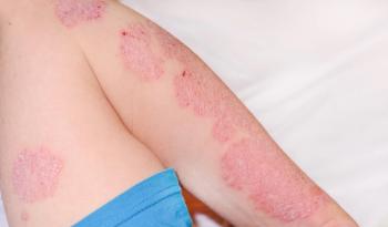
Epidermal AXL Expression Lower in Psoriatic Skin
The findings highlight new data on AXL expression in inflammatory diseases.
To evaluate the expression of the tyrosine kinase receptor AXL in psoriasis, investigators examined the gene expression levels of patients with psoriasis and healthy controls. Ito et al found that epidermal AXL expression was decreased and infiltration of AXL-expressing dendritic cells was increased in psoriasis.1
AXL is involved in many cellular processes and an overexpression of AXL has been seen in a variety of solid tumors and hematologic malignancies. Researchers wanted to better understand AXL expression and its function in non-malignant conditions.
Using quantitative real-time polymerase chain reaction, Ito et al studied the AXL gene expression levels in 7 patients with
AXL-positive cells were seen in the dermis of patients with psoriasis and controls, although the levels were higher in skin with psoriasis.
The expression levels of AXL in epidermal keratinocytes were also evaluated. Findings revealed that normal human epidermal keratinocytes (NHEKs) showed substantial amounts of AXL in homeostasis, and stimulation with the antimicrobial peptide LL37 caused a decrease in AXL expression. Between interleukin-17A-stimulated (IL-17A) and unstimulated NHEKs, AXL expression was comparable. AXL expression was downregulated in psoriatic epidermis and in keratinocytes of LL-37-stimulated psoriatic conditions.
When stimulated with epidermal growth factor (EGF) or transforming growth factor (TGF)-α, HaCaT cells demonstrated a concentration-dependent increase. There was a significant difference at 10 ng/mL of EGF and 5 ng/mL of TGF-α compared to no stimulation. Findings indicated that an increase induced by growth factors in combination with IL-17A was not a result of AXL signaling in HaCaT keratinocytes.
Examining the function of AXL in producing inflammatory factors in epidermal keratinocytes under IL-17A and TLR3 ligand poly (I:C) revealed that the expressions of the genes DEFB4A, IL36G, CXCL8, and CCL20 were significantly induced by IL-17A stimulation.
Ito et al found that when epidermal keratinocytes were stimulated with factors such as poly (I:C), IL-17A, EGF, and TGF-α to activate signaling pathways, cell proliferation and cytokine production were induced, but AXL involvement was not confirmed.
Ito et al concluded that their study “demonstrated a decreased expression of AXL specifically in the epidermis in patients with psoriasis.” They also noted that AXL mRNA expression was lower in psoriatic skin compared to control skin and found an inverse relationship between neutrophil-to-lymphocyte ratio and platelet-to-lymphocyte ratio. Limitations of the study included the small sample size.
Reference
- Ito Y, Shibata S, Koyama A, Li L, Sugimoto E, Taira H, et al. Decreased epidermal AXL expression and increased infiltration of AXL-expressing dendritic cells in psoriasis. J Cutan Immunol Allergy. 2023;00: 1–11.
https://doi.org/10.1002/cia2.12319
Newsletter
Like what you’re reading? Subscribe to Dermatology Times for weekly updates on therapies, innovations, and real-world practice tips.


