|Articles|December 8, 2022
Eccrine Porocarcinoma: A Rare Cutaneous Tumor
Author(s)Fazila Rajab
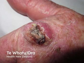
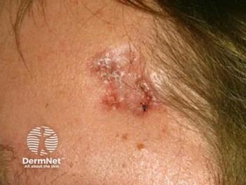
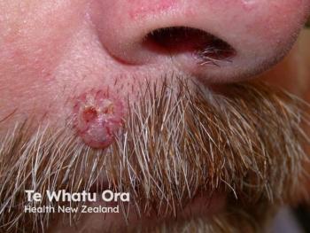
Eccrine porocarcinoma is a rare, aggressive tumor that originates from the intraepidermal ducts of eccrine glands.
Advertisement
Eccrine porocarcinoma (EPC), often known as porocarcinoma, is a rare, aggressive tumor that originates from the intraepidermal ducts of eccrine glands.1 The tumor may typically present as an asymptomatic single dome-shaped nodule ranging from 0.4cm to 20cm in size. The lesion can be erythematous and may later progress to painful ulceration.1
EPC accounts for about 0.005% to 0.01% of all malignant cutaneous tumors and is characterized by highly invasive and metastatic potential, with lymph nodes and lungs being the most common metastatic sites.2-4 The tumor is mainly witnessed in the skin of the upper and lower extremities, head and neck region, and trunk of older adults aged over 60 years.3 The lower extremities are the most commonly affected site in women, whereas men mostly witness the tumor in the head and neck region.3
Epidemiology:
Since EPC is a rare condition, limited research has been conducted to address the epidemiology of the disease. Data collected from SEER-18 shows that EPC is more common in men than women, with the highest incidence rates reported among non-Hispanic Whites.3 Although the tumor primarily affects older adults,3 few studies have also reported EPC in younger patients.1
A study conducted in England reported the overall incidence rate of EPC at 1.9 per 100,000 people in the country, which is at least ten times higher than in other regions.5 Improvements in histological examination may reduce misdiagnoses and increase the number of diagnosed cases in the future.5
Pathogenesis:
The exact cause of EPC is still unknown; however, research revealed that the tumor is most likely to originate from acrosyringium, an intraepidermal duct of the eccrine gland. EPC may emerge either de novo or from a pre-existing benign poroma.1 Moreover, sun exposure and immunosuppression may also play a critical role in EPC tumorigenesis. In rare cases, EPC may also be caused by exposure to certain chemical agents, including benzene glue and poisonous gases.
A recently conducted review published in Cancers highlighted the involvement of specific oncogenic drivers, signaling pathways, and cell-cycle dysregulation in EPC tumorigenesis. According to the review, alterations in genes, including TP53, CDKN2A, EGFR, Rb1, HRAS, and MAPK and PI3K-AKT pathway dysregulation may contribute in the development of EPC.1
Diagnosis:
Due to its rarity and resemblance to basal cell carcinoma (BCC), squamous cell carcinoma (SCC), and seborrheic keratosis and the histological presence of atypical cell types, making an accurate diagnosis of EPC is often challenging to dermatologists.2 Since the tumor mostly appears in older people with no clear clinical features, the focus shifts towards immunohistochemical and histopathological examinations for diagnosis.
Ductal differentiation of poromatous basaloid cells and cytologic atypia with advanced margins are the characteristic histological features of EPC.2 Moreover, evidence revealed squamous differentiation, necrosis and ductal formation as the most common histological features of ECP on hematoxylin and eosin (HE) staining. However, it is challenging to diagnose EPC on HE staining alone.1
Immunohistochemistry, using EMA, CEA, CK-7, and S-100 protein, is another diagnostic tool that further aid in accurate diagnosis by identifying ductal structures.2 Data shows that EMA is more sensitive than CEA in analyzing EPC tumors; however, these tests are insufficient to make a definitive diagnosis as they also highlight the eccrine ducts of SCC.
Furthermore, EPC shows thin irregular vascular patterns on dermatoscopy, and whitish globular structures on the light brown background make an important EPC dermatoscopic feature. Studies show that atypical vascular patterns combined with milky-red globular structures are specific to EPC.1
Treatment Options:
The prognosis of EPC is generally good in the early stages when it can be effectively treated with surgical excision. However, the prognosis of patients with metastatic EPC is grim, with between 5 and 24 months of overall survival rate.2
Surgical Excision:
Currently, no treatment guidelines exist for EPC, and it follows the treatment protocols of sweat gland carcinoma. Mohs micrographic surgery (MMS) or complete circumferential peripheral and deep margin assessment (CCPDMA) is the first line of treatment for patients with EPC.6 However, wide local excision (WLE) can also be performed with a resection margin of at least 2cm from the edge.7
A meta-analysis conducted in 2020 revealed that 92.5% of EPC patients were treated with surgical excision; among these cases, WLE was performed in 76.7% of cases.8 However, the role of sentinel lymph node biopsy in treating EPC is unclear and can be considered depending on the patient's condition and advanced disease risk factors.1
Adjuvant therapy:
The efficacy of radiotherapy and chemotherapy for EPC treatment remains controversial. However, few studies suggest that radical radiotherapy can be given post-surgery to reduce recurrence and control metastasis of tumors.6 A review published in the European Review for Medical and Pharmacological Sciences highlighted the effectiveness of adjuvant radiotherapy in cases with close or positive margins and unfavorable histology.9
Immunotherapy using pembrolizumab can also prove to be an effective novel treatment for metastatic EPC; however, limited evidence exists at present, and further research is needed to establish its efficacy.1
References
- Miyamoto K, Yanagi T, Maeda T, Ujiie H. Diagnosis and management of porocarcinoma. Cancers. 2022; 14(21):5232.
https://doi.org/10.3390/cancers14215232 - Seretis K, Bounas N, Lampri E, Lykoudis EG. Eccrine porocarcinoma of the face is a great imitator with aggressive behavior. Dermatol Pract Concept. 2022;12(2):e2022085. Published 2022 Apr 1. doi:10.5826/dpc.1202a85
- Ragi SD, Moseley I, Ouellette S, Rao B. Epidemiology and survival of eccrine porocarcinoma by sex in the United States: A surveillance, epidemiology, and end results database analysis. Dermatol Surg. 2022. doi:10.1097/DSS.0000000000003652
- Salih AM, Kakamad F, Baba HO, et al. Porocarcinoma; presentation and management, a meta-analysis of 453 cases. Ann Med. 2017;20:74–79. doi: 10.1016/j.amsu.2017.06.027.
- Goon PKC, Gurung P, Levell NJ, et al. Eccrine Porocarcinoma of the Skin is Rising in Incidence in the East of England. Acta Derm Venereol. 2018;98(10):991-992. doi:10.2340/00015555-3000
- Shen J, Pan X, Lu Y, Pan D, Ma Y, Zhan R. A case of eccrine porocarcinoma characterized by a progressive increase in the level of Ki-67 index: case report and review of literature. BMC Surg. 2019;19(1):142. Published 2019 Oct 10. doi:10.1186/s12893-019-0595-4
- Worley B, Owen JL, Barker CA, et al. Evidence-based clinical practice guidelines for microcystic adnexal carcinoma: Informed by a systematic review. JAMA Dermatol. 2019;155(9):1059-1068. doi:10.1001/jamadermatol.2019.1251
- Le NS, Janik S, Liu DT, et al. Eccrine porocarcinoma of the head and neck: Meta-analysis of 120 cases. Head Neck. 2020;42(9):2644-2659. doi:10.1002/hed.26178
- Fionda B, Di Stefani A, Lancellotta V, et al. The role of postoperative radiotherapy in eccrine porocarcinoma: a multidisciplinary systematic review. Eur Rev Med Pharmacol Sci. 2022;26(5):1695-1700. doi:10.26355/eurrev_202203_28238
Newsletter
Like what you’re reading? Subscribe to Dermatology Times for weekly updates on therapies, innovations, and real-world practice tips.
Advertisement
Related Content
Advertisement

Latest CME


Advertisement
Advertisement