
|Articles|January 31, 2013
Diagnosis of PAMS involves targeting syndrome look-alikes
The complexities of diagnosing and managing paraneoplastic autoimmune multiorgan syndrome (PAMS) requires a multidisciplinary, team-oriented approach, an expert says.
Advertisement
Irvine, Calif. - The complexities of diagnosing and managing paraneoplastic autoimmune multiorgan syndrome (PAMS) requires a multidisciplinary, team-oriented approach, an expert says.
Aside from palliative care, though, there’s little physicians can do for the majority of patients with PAMS, says Sergei A. Grando, M.D., Ph.D., D.Sc. He is a professor of dermatology and biological chemistry, University of California-Irvine School of Medicine.
Dr. Grando
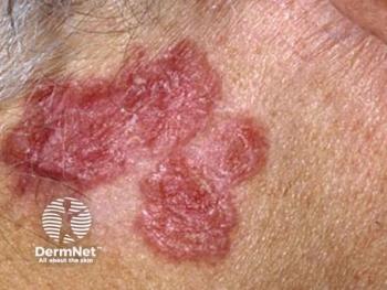
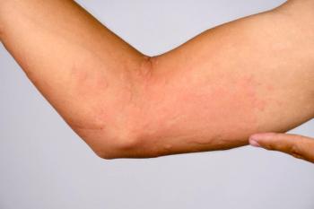
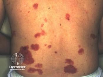
Diagnosing PAMS begins with ruling out look-alike conditions including pemphigus vulgaris, bullous pemphigoid, cicatricial or mucous membrane pemphigoid, erythema multiforme, lichen planus pemphigoides, Stevens-Johnson syndrome, and graft-versus-host disease, he says.
“Other diseases that may look like PAMS include toxic epidermal necrolysis, radiation dermatitis, fungal infection and, because of their oral involvement, some yeast and herpetic infections,” he says.
Physicals for patients with possible PAMS should include lymph node and abdominal exam, Dr. Grando says. He also recommends studying mucocutaneous specimens histologically and via direct immunofluorescence (DIF), and indirect immunofluorescence (IDIF) for serum samples. At this stage, he strongly suggests teaming with experts.
“It would be next to impossible for a non-academic dermatologist not affiliated with a university to do what must be done,” he says. This includes CT scans of the chest, abdomen and pelvis, a complete blood count including serum protein electrophoresis, and diagnostic biopsies from areas of concern, Dr. Grando says. “Clearly, a dermatologist cannot do it alone.”
Diagnostic criteria
Current diagnostic criteria for PAMS include the presence of painful, progressive stomatitis, as well as acantholysis or lichenoid or interface dermatitis (Anhalt GJ. J Investig Dermatol Symp Proc. 2004;9(1):29-33. Review). Previous diagnostic criteria also included confirmation through immunoprecipitation of all four antibodies (those that target proteins weighing 250, 230, 210 and 190 kDa) discovered in the case series from which the criteria were drawn. Dr. Grando says, however, that revised criteria seek only any two of these. “All the patients that my colleagues and I have seen usually have up to three antibodies, but not all four.”
Current published criteria also include proof of an underlying lymphoproliferative neoplasm. “That may not be possible. Only about 70 percent of patients have hematologic malignancies. And remember, one-third of patients have no known malignancies at the time of cutaneous eruption.”
In addition to these criteria, Dr. Grando says physicians must not overlook respiratory findings. In fact, based on published papers and his clinical experience, major criteria for PAMS should include polymorphous mucocutaneous eruption; painful, persistent stomatitis; and respiratory involvement.
Even when asked, he says, “Patients may not tell you whether they have respiratory problems because it would not occur to them to complain to a dermatologist about a cough.” However, he says that in a case series of 28 patients who had PAMS with Castleman’s disease, 26 had respiratory involvement (Nikolskaia OV, Nousari CH, Anhalt GJ. Br J Dermatol. 2003;149(6):1143-1151). Clinicians should be aware during the physical exam that respiratory problems could present before the clinician discovers an underlying neoplasm or cutaneous eruption, he says.
As a fourth major criterion, he says DIF often shows both intercellular and basement membrane linear staining. As in the revised criteria above, he says that immunoprecipitation (which is no longer available in the United States) or immunoblotting must confirm the presence of any two characteristic PAMS antibodies.
In the mucosa, Dr. Grando says, autoantibodies target bronchial epithelial cells, inducing apoptosis. The dead respiratory cells detach from the lamina propria and from neighboring cells and aspirate distally. “They go from the large bronchial tree down and occlude small airways in alveolar sacs.” The entire sloughed layer is cytokeratin 14-positive, he says, although cytokeratin 8-positive cells detach from the alveolar sacs and plug small airways as well.
Other factors
Antibody activity alone does not explain the detachment of respiratory cells, however, according to Dr. Grando.
“We also see mononuclear CD68 cells in the vicinity of sloughed cells,” as well as within the sloughed epithelial sheets. “Clearly, we see factors of both cell-mediated cytotoxicity and humoral cytotoxicity.” To elucidate these mechanisms, Dr. Grando and colleagues used neonatal desmoglein 3 knockout mice. “If you inject them with pemphigus vulgaris antibody, they develop blisters (Nguyen VT, Ndoye A, Bassler KD, et al. ArchDermatol. 2001;137(2):193-206). We wondered what would happen if we inject serum from a patient with PAMS.” The mice developed blisters, but not overnight, as is usually the case with PV serum, Dr. Grando says. Instead, researchers observed a two-stage process: about 24 hours after injection, the skin became erythematous.
“If you do a biopsy, you will see very minor microvesiculation, but most importantly, you can see mononuclear cell infiltrate,” he says. Two days after injection, “You see complete disintegration of the epidermis. The same thing happens with respiratory cells.”
Standard approach
Somewhat similarly, “Rituximab has changed how we treat immunobullous disease,” he says. “It is a chimeric mouse and human monoclonal antibody that targets CD20 phosphoprotein on the surface of free and B lymphocytes, as well as malignant B cells. It basically wipes out all the B cells.”
In two early publications, Dr. Grando says, rituximab showed success against PAMS (Borradori L, Lombardi T, Samson J, et al. Arch Dermatol. 2001;137(3):269-272. Heizmann M, Itin P, Wernli M, et al. Am J Hematol. 2001;66(2):142-144). However, he says it’s possible to have PAMS without antibodies. Subsequent trials by other researchers failed to confirm these findings. “This is not surprising, because rituximab does affect T-cell-mediated response, but apparently not enough to block the immune response entirely.”
Conversely, Dr. Grando says alemtuzumab, which targets the T-cell marker CD52, has achieved dramatic results against PAMS in European research (Hohwy T, Bang K, Steiniche T, et al. Eur J Haematol. 2004 Sep;73(3):206-209).
“You can have PAMS without antibodies. But you cannot have PAMS without autoimmune T cells. If we go in that direction, we may actually improve prognosis. Otherwise, the fatality rate for PAMS is more than 90 percent (Nousari HC, Deterding R, Wojtczack H, et al. N Engl J Med. 1999;340(18):1406-1410),” he says.
Most patients succumb secondary to infection or respiratory failure, Dr. Grando says.
“Usually, it’s a multiorgan failure.” Moreover, one group of researchers has suggested that there are two types of PAMS. In the fatal Group A, patients have distinctive cell-surface antibodies detectable by complement IDIF that are not present in nonfatal Group B (Beutner EH, Pelton S, Hashimoto T, et al. J Am Acad Dermatol. 2002;47(6):841-851). DT
Disclosures: Dr. Grando reports no relevant financial interests.
Advertisement
Related Content
Advertisement


Latest CME


Advertisement
Advertisement
Trending on Dermatology Times
1
KT-621 in Atopic Dermatitis: What Early EASI and Pruritus Data Show
2
Once-Daily Zasocitinib Rivals Injectable Biologics for Skin Clearance, Phase 3 Data Show
3
Filling the HS Treatment Gap: Ruxolitinib Targets Early-Stage Disease
4
What Dermatologists Need to Know About the First New Sunscreen Ingredient in Decades
5