
|Articles|August 1, 2006
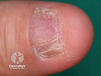
Dermoscopy detects melanoma incognito
San Diego - Using a dermoscope can help dermatologists diagnose melanomas that don't exhibit classic warning signs, says an expert.
Advertisement
San Diego - Using a dermoscope can help dermatologists diagnose melanomas that don't exhibit classic warning signs, says an expert.
"Everyone who does dermoscopy uses it for lesions that look clinically atypical," says Robert Johr, M.D., clinical professor of dermatology and pediatrics (and director of the Pigmented Lesion Clinic) at the University of Miami School of Medicine, and a Boca Raton, Fla.-based private practitioner.
"However," he tells Dermatology Times, "we saw seven cases in which we used dermoscopy on lesions that didn't look particularly worrisome clinically, and to our surprise diagnosed melanomas (study submitted for publication)."
"But if one looks at this type of lesion with dermoscopy," he says, "there might be clues that raise one's index of suspicion enough to warrant a histopathologic diagnosis."
Justification for dermoscopy
Critics of dermoscopy say that if a lesion looks bad, just remove it, Dr. Johr says.
However, he adds, "That is not cutting-edge for a patient with multiple atypically pigmented skin lesions or even a solitary banal-appearing melanoma incognito."
Most high-risk dysplastic nevi or melanoma possess clinical criteria that raise suspicion, he says.
"But we know that melanoma can mimic many benign lesions and nonmelanoma skin cancers," Dr. Johr explains.
In a review of more than 9,000 histopathological specimens of suspected seborrheic keratoses, "A significant number of those turned out to be melanomas (Izikson L, Sober AS, Mihm MC et al. Prevalence of melanoma clinically resembling seborrheic keratosis. Arch Dermatol.2002; 138:1562-1566)," Dr. Johr says.
Strategies from experience
To that end, Dr. Johr and his colleagues offer the following lessons - gleaned from their case series - to prevent missing melanoma incognito:
"But we decided to look at it anyway with dermoscopy. It was a banal-looking, small congenital melanocytic nevus that had areas with the globular pattern that we expected. But to our surprise, we also saw a bluish-white color that is considered a melanoma-specific criterion," Dr. Johr says.
Only because of the presence of the blue-white color, he adds, "A biopsy was performed, and the seemingly banal congenital nevus turned out to be an early invasive melanoma arising in a congenital nevus."
"We had a 44-year-old Caucasian woman who came in for a routine skin examination," Dr. Johr says.
Upon examining a few banal-looking nevi on her trunk with dermoscopy, he says, "Most of the banal-appearing nevi had what we expected to see - regular pigment network and regular dots and globules."
However, one of the lesions had a different dermoscopic appearance. Dr. Johr explains, "It was totally unexpected - we saw foci of bluish color, brown to black globules and whitish lines at the periphery. Because there was not a correlation between what we saw clinically and with dermoscopy, a biopsy was performed, and it turned out to be a level III 0.9 mm melanoma."
A 30-year-old female with a history of melanoma presented for a routine follow-up visit.
Advertisement
Related Content
Advertisement


Latest CME


Advertisement
Advertisement
Trending on Dermatology Times
1
FDA Accepts Addition of Bemotrizinol as First New Sunscreen Ingredient in 20 Years
2
Dual-Action Microneedle Patch Shows Promise for Psoriasis in Preclinical Study
3
AI Analysis Reveals Menopausal Hair Loss Linked to Systemic Health and Inflammation
4
Novel Botanical Moisturizer Demonstrates Superior Efficacy Over Metronidazole in Phase 2 Rosacea Trial
5