

|Articles|July 1, 2016
Dermoscopy can enhance your diagnostic ability and accuracy
The role of dermoscopy has expanded beyond melanoma detection. Experts offer best practices and explain how to embrace and use this powerful tool to get more complete and accurate diagnoses.
Advertisement
Dermoscopy is on the rise among U.S. dermatologists, especially the ones who are newer to the profession and have undergone training in it.
This lesion has a starburst-like pattern. This is a spitzoid melanoma.A 2014 survey found that 98% of dermatologists with five or fewer years of experience said they use dermoscopy compared to 81% of those with 20 or more years.1
There are plenty of reasons to embrace dermoscopy. âWhile dermoscopy was invented to improve diagnosing melanomas and melanoma detection, itâs evolved over the years and can be used in infections, lupus, different types of rashes and inflammatory conditions,â says New York City dermatologist Omar Noor, M.D., FAAD. âEvery day, people are finding new uses for diagnosing skin conditions.â
But dermoscopy can be intimidating at first, especially for dermatologists who didnât encounter it during their training. âItâs an incredible tool, but there is definitely a learning curve and a comfortability curve,â Dr. Noor says. Itâs crucial, he says, to understand that dermoscopy is not a replacement for biopsies, although it does help reduce scarring by giving dermatologists better insight into which lesions actually need to be biopsied. âWhile itâs very helpful in deciding whether something is good or bad, it doesnât take place of histology,â Dr. Noor says.
How can dermatologists embrace dermoscopy if theyâre new to it or need to get better? Dermatology Times asked several top dermatologists for tips and tricks. Hereâs what they said:
Of similar interest:
Next:
Embrace dermoscopyâs powers often
Pedram Gerami, M.D., associate professor of dermatology and pathology with Northwestern University and director of the melanoma program in the Northwestern Skin Cancer Institute, recommends that dermatologists perform dermoscopy on a regular basis in order to improve their skills. âI take dermoscopic images of every lesion that I biopsy and review the dermoscopic image with the histopathologic findings,â Dr. Gerami says. âPerforming dermoscopy on all your patients and correlating with the pathology will help improve your skills quickly.â
Dr. GeramiHe also suggests that dermatologists always keep the clinical exam in mind. âAvoid becoming one-dimensional and ignoring the gross examination,â Dr. Gerami says, adding that âthe power of dermoscopy can be magnified by incorporating clues from the gross clinical exam, so itâs important not to forget that aspect of things.â
Laura Ferris, M.D., an assistant professor with the University of Pittsburghâs Department of Dermatology, offers this advice to Dermatology Times: âDon't start by using dermoscopy to decide what NOT to biopsy. If you are suspicious clinically, but not based on dermoscopy, do a biopsy and take photosâclinical and dermatoscopic. Then go back and see if you were right and what features were present that could have helped you make that diagnosis.â
Pick up on an algorithm
To help dermatologists interpret dermoscopy results, Ashfaq Marghoob, M.D., and colleagues have created a new algorithm called TADA (Triage Amalgamated Dermoscopy Algorithm). Itâs âdesigned for the busy dermatologist or clinician with limited knowledge of dermoscopy,â says Dr. Marghoob, who is attending physician with the dermatology service at Memorial Sloan Kettering Cancer Center and Memorial Sloan Kettering Skin Cancer Center.
Dr. MarghoobThe challenge is that dermatologists tend to disagree over what they see via dermoscopy. However, âwhile our research has shown that while the inter-observer agreement for any specific structure is low, the subjective interpretation of whether dermoscopic structures are distributed in an organized or disorganized manner is much higher,â Dr. Marghoob says.
This distinction is important because it offers insight into whether a lesion is cancerous.
âThe first step in TADA is to eliminate lesions that are clearly benign from entering the algorithm,â Dr. Marghoob says. âThus, lesions that have clear-cut clinical and dermoscopic features of a seborrheic keratosis, angioma or dermatofibroma are excluded.â
The next step is to evaluate all other lesions. âFor this, the observer does not necessarily have to know the significance of any specific structure,â Dr. Marghoob says. âThey simply have to see whether the structures are distributed in an organized or disorganized manner. Most skin cancer will appear disorganized, and most benign lesions will appear organized.â
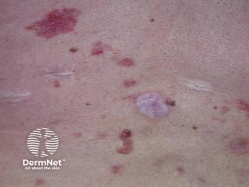
This lesion has a disorganized distribution of colors and structures. Based on TADA this lesion should be biopsied (melanoma 0.35mm).A disorganized lesion should be biopsied. As for the others, âif it is deemed to be organized, then it is evaluated further to ensure that a rare symmetric skin cancer is not missed,â Dr. Marghoob says. âThese include some basal cell carcinomas, keratoacanthomas, amelanotic melanoma, nodular melanoma and spitzoid melanoma.â
As part of this evaluation, he says, âthe observer needs to look for the following: blue-black-gray color, negative network, shiny white structures on polarized light, vessels or ulceration. If any of these are present, then a biopsy should be considered.â
A study about the TADA evaluation system is now in review, Dr. Marghoob says.
Next:
Know what the structures mean
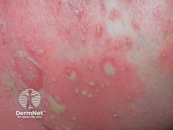
This lesion has an organized distribution of colors and structures but it has a negative network and according to TADA should be biopsied (spitzoid melanoma 0.59mm).Dr. Marghoob offers this advice about other things to watch out for via dermoscopy:
⢠Structures that look like leaves and spoke wheels: âThe presence of these structures is diagnostic of basal cell carcinoma and highly suggestive of the superficial variant,â he says. âIf this is seen, the M.D. could do a shave excision of the lesion.â
⢠Structures arranged in string-of-pearls patterns: These are 100% percent specific for clear cell acanthoma, a rare condition. âIf they see this, then it confirms that the lesion is benign,â he says.
⢠The presence of shiny white blotches and strands within the same lesion: These are highly suggestive of basal cell carcinoma, he says, and the lesion should be biopsied. This is a new finding, reported just this year.2
⢠Angulated zig-zag lines (grayish lines that coalesce creating a zig-zag pattern), which can coalesce into polygonal shapes: âThis is seen in actinic keratosis and melanomas located on sun-damaged skin,â he says. âIf this structure is seen, the dermatologist should palpate the lesion. If the lesion has a rough texture, then it is likely a pigmented actinic keratosis. If the surface is smooth, then it is most likely melanoma.â
He cautions that palpable lesions should never be monitored unless they are clearly benign.
This lesion has an organized pattern but reveals dotted and linear vessels. Based on the rules of TADA this lesion needs to be biopsied (amelanotic melanoma 0.35mm).Disclosures:
Dr. Gerami, Dr. Ferris, Dr. Noor and Dr. Marghoob report no relevant disclosures.
References:
Murzaku EC, Hayan S, Rao BK. Methods and rates of dermoscopy usage: a cross-sectional survey of US dermatologists stratified by years in practice. J Am Acad Dermatol. 2014;71(2):393-5.
Navarrete-Dechent C, Bajaj S, Marchetti MA, Rabinovitz H, Dusza SW, Marghoob AA. Association of shiny white blotches and strands with nonpigmented basal cell carcinoma: evaluation of an additional dermoscopic diagnostic criterion. JAMA Dermatol. 2016 May 1;152(5):546-52
Advertisement
Related Content
Advertisement


Latest CME


Advertisement
Advertisement
Trending on Dermatology Times
1
Dermalogica Enters Medical Aesthetics with FDA-Cleared Microneedling PRO Pen
2
Social Media Mythbusters: Hypochlorous Acid Sprays
3
Quoin Submits First-Ever IND Application for Peeling Skin Syndrome Therapy
4
Topical GX-03 Hits 92.6% EASI-50 at Week 4 in Phase 2 AD Interim
5