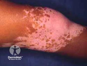
Dermoscopic Monitoring of Vitiligo Lesions Reveal Disease Activity, Evolution Insights
Lesional stability is a necessary requirement for determining the course of treatment for vitiligo.
In a monocenter observational longitudinal study published in Dermatology Practical and Conceptual, researchers delved into the dynamic nature of vitiligo patches, shedding light on their evolution over an 18-month period from April 2021 to October 2022.
The study, conducted at a tertiary hospital, selected 200 vitiligo patches from 60 patients. This research, according to study authors, aimed to provide deeper insights into the behavior of vitiligo lesions over time.
The study included patients of all age groups with vitiligo who were willing to undergo treatment. Exclusion criteria comprised mucosal and scalp vitiligo, lesions less than 1 cm2, and patches previously treated or exhibiting morphological changes
Investigators documented patient history and clinical photographs. Patch size changes were tracked over a period of 6 months using graph paper and transparent sheets. Dermoscopy, performed with a Dinolite video dermoscope, examined predefined parameters such as borders, pigment network, and trichrome pattern. Tailor-made topical/systemic therapies were administered based on patient response.
Of the 200 patches, 183 were studied, with an average patient age of 24.21 years. Female patients exhibited 55.73% of the patches. The study revealed that after 6 months, 64.58% of patches were responsive, 19.28% were progressive, and 11.45% exhibited no change.
Dermoscopic findings reflected significant changes in responsive and progressive patches. Ill-defined borders were observed in both groups. Leukotrichia exhibited reversals in responsive patches. Perifollicular pigmentation and perilesional hyperpigmentation were noted as markers of re-pigmentation and therapy response.
"Our study concluded that a well-defined border is associated with the static nature of a vitiligo patch. However, lesions with an ill-defined border and trichrome pattern suggest the dynamic nature of the patch and are not reliable markers to define the prognosis and instability of the condition," study authors wrote. "Perifollicular pigmentation and perilesional hyperpigmentation are a marker of re-pigmenting vitiligo and can predict the response to therapy. Dermoscopic findings of leukotrichia, satellite lesions and micro-koebnerisation are significantly associated with progressive vitiligo. Leukotrichia may reverse after therapy. Repeated dermoscopic evaluation of lesions in a serial manner to assess disease activity helps understand their evolving nature and is a valuable tool in planning appropriate further treatment."
Reference
Kamath C, Dhurat R, Shah B, Sharma R, Kowe PA, Chamle S. Monitoring of Vitiligo Patches Over Six Months to Validate Dermoscopic Findings of Lesional Stability. Dermatol Pract Concept. 2023;13(4):e2023277. Published 2023 Oct 1.
Newsletter
Like what you’re reading? Subscribe to Dermatology Times for weekly updates on therapies, innovations, and real-world practice tips.


