

|Articles|April 19, 2023
Dermoscopic Features in Melanoma Associated With 3 Genes
Author(s)Emma Andrus, Assistant Editor
Researchers identified several dermoscopic features associated with genes LINC00518, PRAME, and TERT.
Advertisement
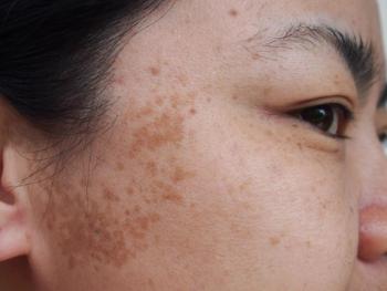
Several dermoscopic features of cutaneous melanoma can be associated with 3 primary genes: LINC00518, PRAME, and TERT.
There are 3-gene expression profiling pigmented lesion assays (3-GEP PLA) which detect the 3 genes in a noninvasive manner. These genes are Long Intergenic Non-Protein Coding RNA 518 (LINC00518), Preferentially Expressed Antigen in Melanoma (PRAME), and mutated Telomerase Reverse Transcriptase (TERT).
In a recent retrospective case-control study,1 researchers sought to identify dermoscopic features unique associated with the 3 genes, citing an absence of literature related to melanoma-associated genes and dermoscopic features.
Between April 2021 and August 2021, researchers collected and analyzed data from 351 unique patients, amassing a total of 510 suspicious pigmented skin lesions. All patients had undergone 3-GEP PLA testing and adhesive patch sample collection. However, 67 samples were deemed insufficient for testing, and 50 cases did not have dermoscopy or clinical images associated with their medical record. These were excluded from analysis, leaving 393 clinical samples remaining.
In lesions with high levels of dermoscopic or clinical concerns, patients underwent physical biopsies.
Experts in dermoscopy were PLA blinded, and they reviewed all biopsy and sample images. Results were categorized as atypical melanocytic nevus, benign lesion, melanoma skin cancer, or non-melanoma skin cancer when applicable. The experts also reviewed the images for the following features:
- Asymmetry (including asymmetry of color or pattern)
- Atypical polymorphous vessels
- Blue-gray dots/peppering
- Blue structures
- Blue-white veil
- Broadened pigment network
- Crystalline structures
- Diameter > 6 mm
- Irregular borders
- Multiple brown dots/globules
- Multiple colors
- Negative pigment network
- Pseudopods/streaks/radial streaming
- Round structures
- Scar-like depigmentation
- Structureless areas >10%
As a result, researchers found that lesions associated with variables such as blue color, a blue-white veil, color asymmetry, and/or scar-like depigmentation were significantly associated with increased odds of PRAME positivity in the univariable analysis.
Furthermore, lesions with blue color, blue-gray dots/peppering, and/or negative pigment networks were significantly associated with increased odds of LINC00518 positivity in the univariable analysis.
Also in the univariable analysis, researchers found that lesions exhibiting greater than 1 of the ABCD criteria, asymmetry, atypical polymorphous vessels, blue color, blue-gray dots/peppering, blue-white veil, crystalline structures, a diameter > 6 mm, and negative pigment network were significantly associated with increased odds of TERT positivity.
These findings were all associated with a comparison of lesions without these characteristics.
One limitation of the study was inter-reader variability of the clinical dermoscopy images. Additionally, researchers cited the high number of “quantity not sufficient” sampled lesions as another potential limitation, going on to recommend further and larger multi-center studies of a similar nature.
“We found that PRAME expression was most significantly associated with dermoscopic color asymmetry, LINC00518 expression was most significantly associated with blue color and negative pigment network, and mutations in the TERT promoter were most significantly associated with the presence of atypical polymorphous vessels in the context of pigmented skin lesions,” study authors wrote. “The information presented suggests a hierarchy in the implications of these features and may guide dermatology providers during their evaluation, decision to use additional ancillary triage tests, and management of pigmented skin lesions.”
Reference
- Ludzik J, Becker AL, Latour E, Lee C, Witkowski A. Dermoscopic features associated with 3‐gep pla: Linc00518, PRAME, and TERT expression in suspicious pigmented lesions. Skin Research and Technology. 2023;29(4). doi:10.1111/srt.13323
Advertisement
Related Content
Advertisement


Latest CME


Advertisement
Advertisement
Trending on Dermatology Times
1
Real-World Study Finds Experienced Dermatologists Outperform AI in Skin Cancer Diagnosis
2
New Consensus Defines Remission, Disease Activity in Atopic Dermatitis
3
Beyond Retinol: Evaluating the Stability, Safety, and Gene-Expression Profile of Bakuchiol Ferulate
4
Zasocitinib Shows High Skin Clearance at Hard-to-Treat Psoriasis Sites
5