Consider rare tumor while making diagnoses
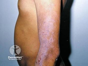
MAC typically presents as a smooth dermal nodule or plaque that displays a flesh-colored or yellow hue.
Chicago - Even though it is considered uncommon among skin carcinomas, dermatologic surgeons should keep microcystic adnexal carcinoma (MAC) in the back of their minds as a possible diagnosis to ensure correct and timely treatment, according to Allison T. Vidimos, R.Ph., M.D., at the American Academy of Dermatology's Academy '05.
"If the clinician doesn't clinically suspect MAC and he/she does not perform a deep enough biopsy, the dermatopathologist won't see the features that are characteristic of the tumor," says Dr. Vidimos, chairman and staff physician, department of dermatology, at the Cleveland Clinic Foundation. "The pathologist may only see the superficial histologic features that can often look benign, and be interpreted as a syringoma or other adnexal neoplasm.
"If a MAC is not treated appropriately the first time and is allowed to recur, this may lead to extensive surgery and possible local or distant metastases," she says.
Because MAC can be very aggressive in its growth pattern, the clinician should recognize the clinical appearance and the most common locations of this tumor. MAC typically presents as a smooth dermal nodule or plaque that displays a flesh-colored or yellow hue. The average size at presentation is 2 cm. This tumor most frequently occurs on the upper lip, cheek and periorbital skin, but has also been found in the axilla and on the scalp, ear, trunk and buttocks. MAC typically occurs in patients ages 50 to 70, but may also occur in children.
Clinically, MAC tumors are often misdiagnosed as basal cell carcinoma, squamous cell carcinoma, cysts, adnexal tumors or scars, Dr. Vidimos explains. Histologically, the tumors are frequently misinterpreted as morpheaform basal cell carcinomas, desmoplastic squamous cell carcinomas, desmoplastic trichoepitheliomas, syringomas, trichoadenomas, eccrine carcinomas or metastatic breast carcinoma. Misdiagnoses are frequently due to inadequate biopsies.
"It is vital to perform a biopsy of adequate depth to aid the dermatopathologist in arriving at the correct diagnosis," Dr. Vidimos says. "A superficial shave biopsy is usually not adequate, and a punch biopsy should go to the subcutaneous fat to demonstrate the overall architecture, invasiveness and characteristic histologic findings."
The clinician should also provide a good clinical description and differential diagnosis list to the pathologist.
Case in point
An autopsy of a 73-year-old woman with a facial MAC present for over 20 years revealed perineural intracranial spread along the optic nerve. Metastases were also found in her clavicle, ribs and liver (Ohta M, Hiramoto M, Ohtsuka H. Dermatol Surg. 2004:957-960).
"MAC tends to spread along nerves, and in this particular case, it actually grew along the optic nerve and spread to the subdural space, as well as via a hematogenous route to distant sites," Dr. Vidimos says. "Interestingly, the subject did not die from the metastatic MAC, but from pneumonia."
Microcystic adnexal carcinomas may display intramuscular, perichondrial and periosteal involvement as well as permeation of vascular adventitia. The tumor also shows "shelving" or "skating" features along fascial or capsular planes, such as muscle, galea, perichondrium or periosteum, with bony invasion and perineural spread. The potential for local invasion was shown in a case of a 51-year-old woman with MAC of the lower lip that extended through her mental and inferior alveolar nerves into the ramus of the mandible, with bone marrow replacement by tumor cells (Birkby CS, Argenyi ZB, Whitaker DC. J Dermatol Surg Oncol. 1989;15:308-312).
Timely, appropriate treatment
While the occurrence of MAC tumors is relatively rare, with only 17 cases seen by Dr. Vidimos and her Mohs surgery colleagues at the Cleveland Clinic from 1987 to 2003, proper treatment remains central to controlling these tumors.
Newsletter
Like what you’re reading? Subscribe to Dermatology Times for weekly updates on therapies, innovations, and real-world practice tips.


