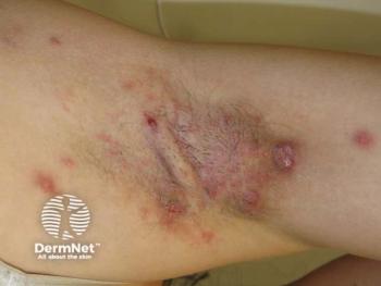
Cameras can be valuable tool for derms
Working with a visual medium is what dermatology is all about.
Orlando - Advancements in digital photography make the use of cameras almost irresistible to dermatologists.
Working with a visual medium is what dermatology is all about. But Frank Joa, principle researcher in skincare testing methods for Procter and Gamble in Cincinnati, Ohio, says there is a difference between photography and the clinical imaging that can be so informative for both physician and patient.
Differences He says photography is controlled by "artistic vision" with each photograph standing on its own merit, while clinical imaging is controlled by standardized methods and the ability to be reproduced.
"The amount of control you need over your system depends on how fine a measurement you're trying to make."
'Too good' He provides an example. "If you want to measure hair color, you can just take a picture. But if you're trying to measure a difference in the chestnut brown color between one person and another, or a change within the same person over a period of a few weeks to see how much it fades, you're going to need more control." The problem is that cameras are almost too good. With the proliferation of digital cameras on the market, Mr. Joa says anyone can go out and buy a "pretty decent" digital camera for $500.
He's heard a number of complaints, however, from researchers who bought cameras and took pictures of samples, or skin cultures, or whatever they were studying but couldn't see any difference in the photos despite differences being noticeable to the naked eye.
"I took one of these cameras and went through the different adjustments and showed them that if you use the automatic settings on these cameras, it will correct out your treatment differences."
Consumers not physicians Mr. Joa covered variables such as exposure, shutter speed, image resolution and white balance. He says cameras are made for consumers not for the clinician, and while almost all of the cameras have automatic settings, only certain cameras have manual settings.
"I compared photos taken using the manual setting, where you basically choose one setting and you take pictures, versus the automatic setting where the camera corrects for changes. If you try to measure a change and the camera corrects for minor changes, you can end up with two images that look the same even though the subjects aren't (the same).
"A camera in fully automatic mode is designed so that if I go into full sunlight and take a picture of the tree I'll be able to see the tree and all the detail. Then, if I go out on a cloudy day and take that same picture, the camera adjusts all the settings so the tree still looks like a tree even though the light level is totally different."
Mr. Joa used the auditorium setting to show the difference in factors that need to be considered when doing clinical imaging.
He compared taking a photo of the entire auditorium to determine how many chairs were filled simply by ensuring that every chair position is visible in the image - to how many people came back to the next seminar by having attendees sit in the same chairs - to determining the eye color of those attending. Many more factors have to be considered in the final photograph: increased image resolution, adjusted lighting to ensure the eye color is illuminated, control for "red eye," checking the visual to make sure eye color is showing (eyes not closed) and to take an image of possible target colors.
Pixels Mr. Joa demonstrated the difference the greater number of pixels makes in the detail of the image. He also explains how the combination of shutter speed and size of aperture can determine how sharp or blurry an image is and how much depth of focus each permits.
He also outlined some of the features physicians should look for in a camera that will be useful for clinical imaging:
Dr. Joa says it's most important for doctors to remember if they want to keep their photo comparisons true, they cannot let the automatic settings on the camera overcompensate for the setting, such as lighting, thereby reducing the visible differences resulting from treatment.
Newsletter
Like what you’re reading? Subscribe to Dermatology Times for weekly updates on therapies, innovations, and real-world practice tips.


