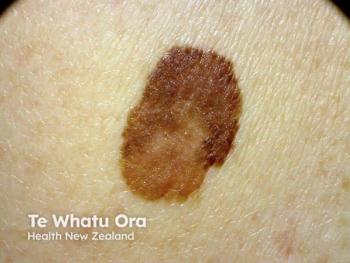
Ablative fractional laser resurfacing associated with pseudomelanoma
The occurrence of several cases of pseudomelanoma after ablative fractional CO2 laser resurfacing underscore the importance of careful pre- and postoperative evaluation to avoid misdiagnosing a benign lesion as malignant melanoma, says Deborah Sarnoff, M.D.
Key Points
New York - The occurrence of several cases of pseudomelanoma after ablative fractional CO2 laser resurfacing underscore the importance of careful pre- and postoperative evaluation to avoid misdiagnosing a benign lesion as malignant melanoma, says Deborah Sarnoff, M.D.
All lesions were biopsied, and the dermatopathologist's report diagnosed melanoma in situ or early evolving melanoma in situ. The first patient underwent wide, deep surgical excision of the lesion that resulted in a facial scar. Subsequent review of the histology in conjunction with the history resulted in revised diagnosis of pseudomelanoma.
Dr. Sarnoff has proposed a mechanism to explain why pseudomelanoma is more likely to occur after fractional CO2 laser resurfacing than following a fully ablative procedure, and she suggests measures for pre- and postoperative evaluation to enable accurate diagnosis.
"Accurate diagnosis will be facilitated by considering the history and clinical presentation carefully," says Dr. Sarnoff, who is also senior vice president of The Skin Cancer Foundation and director of dermatological surgery at her private practice, Cosmetique Dermatology, Laser and Plastic Surgery, New York and Long Island. "For distinguishing pseudomelanoma from malignant melanoma, looking through the retroscope may provide information that is more valuable than that obtained using the dermascope, microscope or special histological stains."
Pseudomelanoma, defined as the reappearance of pigmentation at a site where a melanocytic nevus (usually of an intradermal type) was previously removed, was first described by Ackerman and Kornberg (Arch Dermatol. 1975;111(12):1588-1590), who reported its occurrence in young adults within a few weeks after partial shave excision of an intradermal melanocytic nevus. In addition to appearing after incomplete excision of benign nevi, the condition has been reported to develop after trauma, cryotherapy, dermabrasion and other laser treatments.
"Histology reveals pagetoid spread of atypical melanocytes with nests in all levels of the epidermis overlying an area of fibrosis in the papillary dermis in association with a nevus in the dermis below," Dr. Sarnoff says. "With these features, it can be a challenge for even the most experienced dermatopathologist to differentiate real melanoma from pseudomelanoma, and special stains don't help."
Dr. Sarnoff says that because fractional resurfacing delivers microthermal zones of heat in vertical columns, only a fraction of an existing intradermal nevus will be ablated. Activity of melanocytic cells in areas left intact can be stimulated by the thermal insult, with migration of dermal pigment into the epidermis promoted by post-treatment neocollagenesis and wound-healing responses.
The 112 consecutive patients included in the retrospective review were predominantly women but represented a broad age range and Fitzpatrick skin types I-IV. The three patients who developed a pseudomelanoma ranged in age from 54 to 81 years. All had been treated with fairly aggressive laser settings. Time to appearance of the pigmented lesion was two to three months after the laser resurfacing.
Preventing misdiagnosis
Dr. Sarnoff recommends careful preoperative evaluation to identify any skin-colored nevi, taking care to spread the skin between rhytids to inspect for any lesion that may be concealed between wrinkle crevices. Obtaining excellent baseline digital photographs enables preoperative evaluation by allowing magnification of the images and aids pre- and postoperative comparisons.
She advises evaluating any questionable pigmented nevus prior to the laser procedure with shave biopsy and marking existing benign intradermal nevi prior to resurfacing so that the lesions can be avoided during ablation.
Dr. Sarnoff says the appearance of a melanocytic nevus within one to six months after ablative fractional resurfacing or of multiple new pigmented lesions favors the diagnosis of pseudomelanoma.
Disclosures: Dr. Sarnoff is an investigator for Cynosure and DEKA.
Newsletter
Like what you’re reading? Subscribe to Dermatology Times for weekly updates on therapies, innovations, and real-world practice tips.


